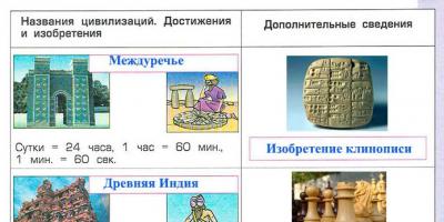
Genetic code– a unified system for recording hereditary information in nucleic acid molecules in the form of a nucleotide sequence. The genetic code is based on the use of an alphabet consisting of only four letters A, T, C, G, corresponding to DNA nucleotides. There are 20 types of amino acids in total. Of the 64 codons, three - UAA, UAG, UGA - do not code for amino acids; they were called nonsense codons and serve as punctuation marks. Codon (encoding trinucleotide) is a unit of genetic code, a trio of nucleotide residues (triplet) in DNA or RNA, encoding the inclusion of one amino acid. The genes themselves do not take part in protein synthesis. The mediator between gene and protein is mRNA. The structure of the genetic code is characterized by the fact that it is triplet, that is, it consists of triplets (triples) of nitrogenous DNA bases, called codons. Out of 64
Properties of the gene. code
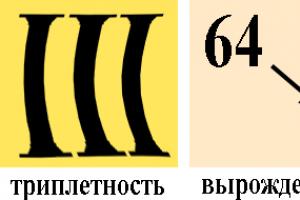
1) Triplety: one amino acid is encoded by three nucleotides. These 3 nucleotides in DNA
are called triplet, in mRNA - codon, in tRNA - anticodon.
2) Redundancy (degeneracy): there are only 20 amino acids, and there are 61 triplets encoding amino acids, so each amino acid is encoded by several triplets.
3) Uniqueness: each triplet (codon) encodes only one amino acid.
4) Universality: the genetic code is the same for all living organisms on Earth.
5.) continuity and indisputability of codons during reading. This means that the nucleotide sequence is read triplet by triplet without gaps, and adjacent triplets do not overlap each other.
88. Heredity and variability are fundamental properties of living things. Darwin's understanding of the phenomena of heredity and variability.
Heredity call the general property of all organisms to preserve and transmit characteristics from parent to offspring. Heredity- this is the property of organisms to reproduce in generations a similar type of metabolism that has developed during the historical development of the species and manifests itself under certain environmental conditions.
Variability is the process of the emergence of qualitative differences between individuals of the same species, which is expressed either in a change under the influence of the external environment of only one phenotype, or in genetically determined hereditary variations resulting from combinations, recombinations and mutations that take place in a number of successive generations and populations.
Darwin's understanding of heredity and variability.
Under heredity Darwin understood the ability of organisms to preserve their species, varietal and individual characteristics in their offspring. This feature was well known and represented hereditary variation. Darwin analyzed in detail the importance of heredity in the evolutionary process. He drew attention to cases of same-suit hybrids of the first generation and splitting of characters in the second generation; he was aware of heredity associated with sex, hybrid atavisms and a number of other phenomena of heredity.
Variability. When comparing many breeds of animals and varieties of plants, Darwin noticed that within any species of animals and plants, and in culture, within any variety and breed there are no identical individuals. Darwin concluded that variability is inherent in all animals and plants.
Analyzing the material on the variability of animals, the scientist noticed that any change in living conditions is enough to cause variability. Thus, Darwin understood variability as the ability of organisms to acquire new characteristics under the influence of environmental conditions. He distinguished the following forms of variability:
Specific (group) variability(now called modification) - a similar change in all individuals of the offspring in one direction due to the influence of certain conditions. Certain changes tend to be non-hereditary.
Uncertain individual variability(now called genotypic) - the appearance of various minor differences in individuals of the same species, variety, breed, by which, existing in similar conditions, one individual differs from others. Such multidirectional variability is a consequence of the uncertain influence of living conditions on each individual.
Correlative(or relative) variability. Darwin understood the organism as an integral system, the individual parts of which are closely interconnected. Therefore, a change in the structure or function of one part often causes a change in another or others. An example of such variability is the relationship between the development of a functioning muscle and the formation of a ridge on the bone to which it is attached. Many wading birds have a correlation between neck length and limb length: birds with long necks also have long limbs.
Compensatory variability consists in the fact that the development of some organs or functions is often the cause of the inhibition of others, that is, there is an inverse correlation, for example, between milk production and meatiness of livestock.
89. Modification variability. Norm of reaction of genetically determined traits. Phenocopies.
Phenotypic variability covers changes in the state of the characteristics themselves that occur under the influence of developmental conditions or environmental factors. The range of modification variability is limited by the reaction norm. A specific modification change in a trait that has arisen is not inherited, but the range of modification variability is determined by heredity. Hereditary material is not involved in the change.
Norm of reaction is the limit of modification variability of a trait. It is the reaction norm that is inherited, not the modifications themselves, i.e. the ability to develop a trait, and the form of its manifestation depends on environmental conditions. The reaction norm is a specific quantitative and qualitative characteristic of the genotype. There are signs with a wide reaction norm, a narrow () and an unambiguous norm. Norm of reaction has limits or boundaries for each biological species (lower and upper) - for example, increased feeding will lead to an increase in the weight of the animal, but it will be within the normal reaction range characteristic of a given species or breed. The reaction rate is genetically determined and inherited. For different traits, the reaction norm limits vary greatly. For example, wide limits of the reaction norm are the value of milk yield, cereal productivity and many other quantitative characteristics, narrow limits are the color intensity of most animals and many other qualitative characteristics. Under the influence of some harmful factors that a person does not encounter in the process of evolution, the possibility of modification variability that determines reaction norms is excluded.
Phenocopies- changes in phenotype under the influence of unfavorable environmental factors, similar in manifestation to mutations. The resulting phenotypic modifications are not inherited. It has been established that the occurrence of phenocopies is associated with the influence of external conditions on a certain limited stage of development. Moreover, the same agent, depending on which phase it acts on, can copy different mutations, or one stage reacts to one agent, another to another. Different agents can be used to induce the same phenocopy, indicating that there is no connection between the result of the change and the influencing factor. The most complex genetic developmental disorders are relatively easy to reproduce, while copying traits is much more difficult.
90. Adaptive nature of modification. The role of heredity and environment in human development, training and education.
Modification variability corresponds to living conditions and is adaptive in nature. Characteristics such as the growth of plants and animals, their weight, color, etc. are subject to modification variability. The occurrence of modification changes is due to the fact that environmental conditions affect the enzymatic reactions occurring in the developing organism and, to a certain extent, change its course.
Since the phenotypic manifestation of hereditary information can be modified by environmental conditions, the organism’s genotype is programmed only with the possibility of their formation within certain limits, called the reaction norm. The reaction norm represents the limits of modification variability of a trait allowed for a given genotype.
The degree of expression of a trait when a genotype is realized under different conditions is called expressivity. It is associated with the variability of the trait within the reaction norm.
The same trait may appear in some organisms and be absent in others that have the same gene. A quantitative measure of the phenotypic expression of a gene is called penetrance.
Expressivity and penetrance are maintained by natural selection. Both patterns must be kept in mind when studying heredity in humans. By changing environmental conditions, penetrance and expressivity can be influenced. The fact that the same genotype can be the source of the development of different phenotypes is of significant importance for medicine. This means that the burden does not necessarily have to manifest itself. Much depends on the conditions in which a person finds himself. In some cases, diseases as a phenotypic manifestation of hereditary information can be prevented by following a diet or taking medications. The implementation of hereditary information depends on the environment. Formed on the basis of a historically established genotype, modifications are usually adaptive in nature, since they are always the result of responses of a developing organism to environmental factors affecting it. The nature of mutational changes is different: they are the result of changes in the structure of the DNA molecule, which causes a disruption in the previously established process of protein synthesis. When mice are kept at elevated temperatures, they produce offspring with elongated tails and enlarged ears. This modification is adaptive in nature, since the protruding parts (tail and ears) play a thermoregulatory role in the body: increasing their surface allows for increased heat transfer.
The genetic potential of a person is limited in time, and quite strictly. If you miss the deadline for early socialization, it will fade away before it has time to be realized. A striking example of this statement are the numerous cases when infants, by force of circumstances, ended up in the jungle and spent several years among animals. After their return to the human community, they could no longer fully catch up with what they had lost: master speech, acquire quite complex skills of human activity, their mental functions of a person developed poorly. This is evidence that the characteristic features of human behavior and activity are acquired only through social inheritance, only through the transmission of a social program in the process of upbringing and training.
Identical genotypes (in identical twins), when placed in different environments, can produce different phenotypes. Taking into account all the influencing factors, the human phenotype can be represented as consisting of several elements.
These include: biological inclinations encoded in genes; environment (social and natural); individual activity; mind (consciousness, thinking).
The interaction of heredity and environment in human development plays an important role throughout his life. But it acquires particular importance during the periods of formation of the body: embryonic, breast, childhood, adolescence and youth. It is at this time that an intensive process of development of the body and formation of personality is observed.
Heredity determines what an organism can become, but a person develops under the simultaneous influence of both factors - heredity and environment. Today it is becoming generally accepted that human adaptation is carried out under the influence of two programs of heredity: biological and social. All signs and properties of any individual are the result of the interaction of his genotype and environment. Therefore, each person is both a part of nature and a product of social development.
91. Combinative variability. The importance of combinative variability in ensuring the genotypic diversity of people: Marriage systems. Medical and genetic aspects of the family.
Combinative variability associated with obtaining new combinations of genes in the genotype. This is achieved as a result of three processes: a) independent chromosome segregation during meiosis; b) their random combination during fertilization; c) gene recombination due to Crossing Over. The hereditary factors (genes) themselves do not change, but their new combinations arise, which leads to the appearance of organisms with different genotypic and phenotypic properties. Thanks to combinative variability a variety of genotypes is created in the offspring, which is of great importance for the evolutionary process due to the fact that: 1)
the diversity of material for the evolutionary process increases without reducing the viability of individuals; 2)
the ability of organisms to adapt to changing environmental conditions expands and thereby ensures the survival of a group of organisms (population, species) as a whole
The composition and frequency of alleles in people and populations largely depend on the types of marriages. In this regard, the study of types of marriages and their medical and genetic consequences is important.
Marriages can be: selective, indiscriminate.
To the non-selective include panmix marriages. Panmixia(Greek nixis - mixture) - step marriages between people with different genotypes.
Selective marriages: 1.Outbreeding– marriages between people who are not related by a previously known genotype, 2.Inbreeding- marriages between relatives, 3.Positively assortative– marriages between individuals with similar phenotypes (deaf-mute, short with short, tall with tall, feeble-minded with feeble-minded, etc.). 4.Negative assortative-marriages between people with dissimilar phenotypes (deaf-mute - normal; short - tall; normal - with freckles, etc.). 4.Incest– marriages between close relatives (between brother and sister).
Inbred and incestuous marriages are illegal in many countries. Unfortunately, there are regions with a high frequency of inbred marriages. Until recently, the frequency of inbred marriages in some regions of Central Asia reached 13-15%.
Medical and genetic significance inbred marriages are very negative. In such marriages, homozygotization is observed, and the frequency of autosomal recessive diseases increases by 1.5-2 times. Inbred populations experience inbreeding depression, i.e. the frequency of unfavorable recessive alleles increases sharply, and child mortality increases. Positively assortative marriages also lead to similar phenomena. Outbreeding has positive genetic benefits. In such marriages, heterozygotization is observed.
92. Mutational variability, classification of mutations according to the level of change in the damage to the hereditary material. Mutations in germ and somatic cells.
Mutation is called a change caused by the reorganization of reproductive structures, a change in its genetic apparatus. Mutations occur spasmodically and are inherited. Depending on the level of change in the hereditary material, all mutations are divided into genetic, chromosomal And genomic.
Gene mutations, or transgenations, affect the structure of the gene itself. Mutations can change sections of the DNA molecule of varying lengths. The smallest region, the change of which leads to the appearance of a mutation, is called a muton. It can only be made up of a pair of nucleotides. A change in the sequence of nucleotides in DNA causes a change in the sequence of triplets and, ultimately, the protein synthesis program. It should be remembered that disturbances in the DNA structure lead to mutations only when repair is not carried out.
Chromosomal mutations, chromosomal rearrangements or aberrations consist of a change in the amount or redistribution of the hereditary material of chromosomes.
Perestroikas are divided into intrachromosomal And interchromosomal. Intrachromosomal rearrangements consist of the loss of part of a chromosome (deletion), doubling or multiplication of some of its sections (duplication), and rotation of a chromosome fragment by 180° with a change in the sequence of gene location (inversion).
Genomic mutations associated with changes in the number of chromosomes. Genomic mutations include aneuploidy, haploidy and polyploidy.
Aneuploidy called a change in the number of individual chromosomes - the absence (monosomy) or the presence of additional (trisomy, tetrasomy, generally polysomy) chromosomes, i.e. an unbalanced chromosome set. Cells with an altered number of chromosomes appear as a result of disturbances in the process of mitosis or meiosis, and therefore a distinction is made between mitotic and meiotic aneuploidy. A multiple decrease in the number of chromosome sets of somatic cells compared to diploid is called haploidy. A multiple increase in the number of chromosome sets of somatic cells compared to diploid is called polyploidy.
The listed types of mutations occur in both germ cells and somatic cells. Mutations that occur in germ cells are called generative. They are passed on to subsequent generations.
Mutations that occur in bodily cells at one or another stage of the individual development of the organism are called somatic. Such mutations are inherited only by the descendants of the cell in which it occurred.
93. Gene mutations, molecular mechanisms of occurrence, frequency of mutations in nature. Biological antimutation mechanisms.
Modern genetics emphasizes that gene mutations consist in changing the chemical structure of genes. Specifically, gene mutations are substitutions, insertions, deletions, and losses of nucleotide pairs. The smallest section of a DNA molecule whose change leads to mutation is called a muton. It is equal to one pair of nucleotides.
There are several classifications of gene mutations . Spontaneous(spontaneous) is a mutation that occurs without direct connection with any physical or chemical environmental factor.
If mutations are caused intentionally, by influencing the body by factors of a known nature, they are called induced. The agent that induces mutations is called mutagen.
The nature of mutagens is diverse- these are physical factors, chemical compounds. The mutagenic effect of some biological objects - viruses, protozoa, helminths - when they penetrate the human body has been established.
As a result of dominant and recessive mutations, dominant and recessive altered traits appear in the phenotype. Dominant mutations appear in the phenotype already in the first generation. Recessive mutations are hidden in heterozygotes from the action of natural selection, so they accumulate in large numbers in the gene pools of species.
An indicator of the intensity of the mutation process is the mutation frequency, which is calculated on average per genome or separately for specific loci. The average mutation frequency is comparable in a wide range of living beings (from bacteria to humans) and does not depend on the level and type of morphophysiological organization. It is equal to 10 -4 - 10 -6 mutations per 1 locus per generation.
Antimutation mechanisms.
A protective factor against the adverse consequences of gene mutations is the pairing of chromosomes in the diploid karyotype of somatic eukaryotic cells. The pairing of alley genes prevents the phenotypic manifestation of mutations if they are recessive.
The phenomenon of extracopying genes encoding vital macromolecules contributes to reducing the harmful consequences of gene mutations. For example, the genes of rRNA, tRNA, histone proteins, without which the life of any cell is impossible.
The listed mechanisms contribute to the preservation of genes selected during evolution and at the same time the accumulation of different alleles in the gene pool of a population, forming a reserve of hereditary variability.
94. Genomic mutations: polyploidy, haploidy, heteroploidy. Mechanisms of their occurrence.
Genomic mutations are associated with changes in the number of chromosomes. Genomic mutations include heteroploidy, haploidy And polyploidy.
Polyploidy– an increase in the diploid number of chromosomes by adding entire chromosome sets as a result of disruption of meiosis.
In polyploid forms, there is an increase in the number of chromosomes, a multiple of the haploid set: 3n – triploid; 4n – tetraploid, 5n – pentaploid, etc.
Polyploid forms are phenotypically different from diploid ones: along with a change in the number of chromosomes, hereditary properties also change. In polyploids, the cells are usually large; sometimes the plants are gigantic in size.
Forms resulting from the multiplication of chromosomes of one genome are called autoploid. However, another form of polyploidy is also known - alloploidy, in which the number of chromosomes of two different genomes is multiplied.
A multiple decrease in the number of chromosome sets of somatic cells compared to diploid is called haploidy. Haploid organisms in natural habitats are found mainly among plants, including higher ones (datura, wheat, corn). The cells of such organisms have one chromosome of each homologous pair, so all recessive alleles are manifested in the phenotype. This explains the reduced viability of haploids.
Heteroploidy. As a result of disturbances in mitosis and meiosis, the number of chromosomes may change and not become a multiple of the haploid set. The phenomenon when one of the chromosomes, instead of being a pair, ends up in a triple number, is called trisomy. If trisomy is observed on one chromosome, then such an organism is called trisomic and its chromosome set is 2n+1. Trisomy can be on any of the chromosomes or even on several. With Double trisomy, it has a chromosome set of 2n+2, triple trisomy – 2n+3, etc.
The opposite phenomenon trisomy, i.e. the loss of one chromosome from a pair in a diploid set is called monosomy, the organism is monosomic; its genotypic formula is 2n-1. In the absence of two different chromosomes, the organism is double monosomic with the genotypic formula 2n-2, etc.
From what has been said it is clear that aneuploidy, i.e. a violation of the normal number of chromosomes leads to changes in the structure and a decrease in the viability of the organism. The greater the disturbance, the lower the viability. In humans, disruption of a balanced set of chromosomes leads to painful conditions known collectively as chromosomal diseases.
Mechanism of occurrence genomic mutations are associated with the pathology of disruption of normal chromosome segregation in meiosis, resulting in the formation of abnormal gametes, which leads to mutation. Changes in the body are associated with the presence of genetically heterogeneous cells.
95. Methods for studying human heredity. Genealogical and twin methods, their significance for medicine.
The main methods for studying human heredity are genealogical, twin, population-statistical, dermatoglyphics method, cytogenetic, biochemical, somatic cell genetics method, modeling method
Genealogical method. This method is based on the compilation and analysis of pedigrees. A pedigree is a diagram that shows the connections between family members. By analyzing pedigrees, they study any normal or (more often) pathological trait in generations of people who are related.
Genealogical methods are used to determine the hereditary or non-hereditary nature of a trait, dominance or recessivity, chromosome mapping, sex linkage, and to study the mutation process. As a rule, the genealogical method forms the basis for conclusions in medical genetic counseling.
When compiling pedigrees, standard notations are used. The person with whom the study begins is the proband. The descendant of a married couple is called a sibling, siblings are called siblings, cousins are called first cousins, etc. Descendants who have a common mother (but different fathers) are called consanguineous, and descendants who have a common father (but different mothers) are called half-blooded; if a family has children from different marriages, and they do not have common ancestors (for example, a child from the mother’s first marriage and a child from the father’s first marriage), then they are called step-children.
Using the genealogical method, the hereditary nature of the trait being studied can be established, as well as the type of its inheritance. When analyzing pedigrees for several characteristics, the linked nature of their inheritance can be revealed, which is used in the compilation of chromosomal maps. This method allows you to study the intensity of the mutation process, assess the expressivity and penetrance of the allele.
Twin method. It consists of studying the patterns of inheritance of traits in pairs of identical and fraternal twins. Twins are two or more children conceived and born by the same mother almost simultaneously. There are identical and fraternal twins.
Identical (monozygotic, identical) twins occur at the earliest stages of zygote fragmentation, when two or four blastomeres retain the ability to develop into a full-fledged organism when separated. Because the zygote divides by mitosis, the genotypes of identical twins are, at least initially, completely identical. Identical twins are always the same sex and share the same placenta during fetal development.
Fraternal (dizygotic, non-identical) occur when two or more simultaneously matured eggs are fertilized. Thus, they share about 50% of their genes. In other words, they are similar to ordinary brothers and sisters in their genetic constitution and can be either same-sex or opposite-sex.
By comparing identical and fraternal twins raised in the same environment, conclusions can be drawn about the role of genes in the development of traits.
The twin method allows you to make informed conclusions about the heritability of traits: the role of heredity, environment and random factors in determining certain human traits
Prevention and diagnosis of hereditary pathology
Currently, the prevention of hereditary pathology is carried out at four levels: 1) pregametic; 2) prezygotic; 3) prenatal; 4) neonatal.
1.) Pregametic level
Carried out:
1. Sanitary control over production - eliminating the influence of mutagens on the body.
2. Liberation of women of childbearing age from work in hazardous industries.
3.Creation of lists of hereditary diseases that are common in a certain area
territories with def. frequent.
2.Prezygotic level
The most important element of this level of prevention is medical genetic counseling (MGC) of the population, informing the family about the degree of possible risk of having a child with a hereditary pathology and providing assistance in making the right decision about childbearing.
Prenatal level
It consists of carrying out prenatal (prenatal) diagnostics.
Prenatal diagnosis– this is a set of measures that is carried out with the aim of determining hereditary pathology in the fetus and terminating this pregnancy. Prenatal diagnostic methods include:
1. Ultrasound scanning (USS).
2. Fetoscopy– a method of visual observation of the fetus in the uterine cavity through an elastic probe equipped with an optical system.
3. Chorionic villus biopsy. The method is based on taking chorionic villi, culturing cells and studying them using cytogenetic, biochemical and molecular genetic methods.
4. Amniocentesis– puncture of the amniotic sac through the abdominal wall and collection
amniotic fluid. It contains fetal cells that can be examined
cytogenetically or biochemically, depending on the expected pathology of the fetus.
5. Cordocentesis- puncture of the umbilical cord vessels and collection of fetal blood. Fetal lymphocytes
cultivated and subjected to research.
4.Neonatal level
At the fourth level, newborns are screened to identify autosomal recessive metabolic diseases in the preclinical stage, when timely treatment begins to ensure normal mental and physical development of children.
Principles of treatment of hereditary diseases
The following types of treatment are available:.
1. Symptomatic(impact on disease symptoms).
2. Pathogenetic(impact on the mechanisms of disease development).
Symptomatic and pathogenetic treatment does not eliminate the causes of the disease, because does not liquidate
genetic defect.
The following techniques can be used in symptomatic and pathogenetic treatment.
· Correction developmental defects using surgical methods (syndactyly, polydactyly,
cleft lip...
· Replacement therapy, the meaning of which is to introduce into the body
missing or insufficient biochemical substrates.
· Metabolism induction– introduction into the body of substances that enhance synthesis
some enzymes and, therefore, speed up processes.
· Metabolism inhibition– introduction into the body of drugs that bind and remove
abnormal metabolic products.
· Diet therapy ( therapeutic nutrition) - elimination from the diet of substances that
cannot be absorbed by the body.
Prospects: In the near future, genetics will develop rapidly, although it is still
very widespread in agricultural crops (breeding, cloning),
medicine (medical genetics, genetics of microorganisms). In the future, scientists hope
use genetics to eliminate defective genes and eradicate diseases transmitted
by inheritance, to be able to treat such serious diseases as cancer, viral
infections.
With all the shortcomings of the modern assessment of the radiogenetic effect, there is no doubt about the seriousness of the genetic consequences awaiting humanity in the event of an uncontrolled increase in the radioactive background in the environment. The danger of further testing of atomic and hydrogen weapons is obvious.
At the same time, the use of atomic energy in genetics and selection makes it possible to create new methods for controlling the heredity of plants, animals and microorganisms, and to better understand the processes of genetic adaptation of organisms. In connection with human flights into outer space, there is a need to study the influence of the cosmic reaction on living organisms.
98. Cytogenetic method for diagnosing human chromosomal disorders. Amniocentesis. Karyotype and idiogram of human chromosomes. Biochemical method.
The cytogenetic method involves studying chromosomes using a microscope. Most often, the object of study is mitotic (metaphase), less often meiotic (prophase and metaphase) chromosomes. Cytogenetic methods are used to study the karyotypes of individual individuals
Obtaining material from an organism developing in utero is carried out in different ways. One of them is amniocentesis, with the help of which, at 15-16 weeks of pregnancy, amniotic fluid is obtained, containing waste products of the fetus and cells of its skin and mucous membranes
The material taken during amniocentesis is used for biochemical, cytogenetic and molecular chemical studies. Cytogenetic methods determine the sex of the fetus and identify chromosomal and genomic mutations. The study of amniotic fluid and fetal cells using biochemical methods makes it possible to detect a defect in the protein products of genes, but does not make it possible to determine the localization of mutations in the structural or regulatory part of the genome. The use of DNA probes plays an important role in identifying hereditary diseases and precise localization of damage to the fetal hereditary material.
Currently, amniocentesis is used to diagnose all chromosomal abnormalities, over 60 hereditary metabolic diseases, and incompatibility of mother and fetus with erythrocyte antigens.
The diploid set of chromosomes of a cell, characterized by their number, size and shape, is called karyotype. A normal human karyotype includes 46 chromosomes, or 23 pairs: 22 pairs of autosomes and one pair of sex chromosomes
In order to make it easier to understand the complex complex of chromosomes that makes up the karyotype, they are arranged in the form idiograms. IN idiogram chromosomes are arranged in pairs in order of decreasing size, with the exception of sex chromosomes. The largest pair is assigned No. 1, the smallest - No. 22. Identification of chromosomes only by size encounters great difficulties: a number of chromosomes have similar sizes. However, recently, through the use of various types of dyes, a clear differentiation of human chromosomes according to their length into bands that can be dyed using special methods and those that cannot be dyed has been established. The ability to accurately differentiate chromosomes is of great importance for medical genetics, as it allows one to accurately determine the nature of abnormalities in a person’s karyotype.
Biochemical method
99. Human karyotype and idiogram. Characteristics of a normal human karyotype
and pathology.
Karyotype- a set of characteristics (number, size, shape, etc.) of the complete set of chromosomes,
inherent in the cells of a given biological species (species karyotype), of a given organism
(individual karyotype) or line (clone) of cells.
To determine the karyotype, a microphotograph or sketch of chromosomes is used during microscopy of dividing cells.
Each person has 46 chromosomes, two of which are sex chromosomes. A woman has two X chromosomes
(karyotype: 46, XX), and men have one X chromosome and the other Y (karyotype: 46, XY). Study
Karyotyping is carried out using a method called cytogenetics.
Idiogram- a schematic representation of the haploid set of chromosomes of an organism, which
placed in a row in accordance with their sizes, in pairs in descending order of their sizes. An exception is made for sex chromosomes, which are especially distinguished.
Examples of the most common chromosomal pathologies.
Down syndrome is a trisomy of the 21st pair of chromosomes.
Edwards syndrome is trisomy on the 18th pair of chromosomes.
Patau syndrome is a trisomy of the 13th pair of chromosomes.
Klinefelter syndrome is a polysomy of the X chromosome in boys.
100. The importance of genetics for medicine. Cytogenetic, biochemical, population-statistical methods for studying human heredity.
The role of genetics in human life is very important. It is implemented with the help of medical genetic counseling. Medical genetic counseling is designed to save humanity from suffering associated with hereditary (genetic) diseases. The main goals of medical genetic counseling are to establish the role of the genotype in the development of this disease and predict the risk of having sick offspring. Recommendations given in medical genetic consultations regarding marriage or prognosis of the genetic usefulness of offspring are aimed at ensuring that they are taken into account by the persons being consulted, who voluntarily make the appropriate decision.
Cytogenetic (karyotypic) method. The cytogenetic method involves studying chromosomes using a microscope. Most often, the object of study is mitotic (metaphase), less often meiotic (prophase and metaphase) chromosomes. This method is also used to study sex chromatin ( Barr bodies) Cytogenetic methods are used to study the karyotypes of individual individuals
The use of the cytogenetic method allows not only to study the normal morphology of chromosomes and the karyotype as a whole, to determine the genetic sex of the organism, but, most importantly, to diagnose various chromosomal diseases associated with changes in the number of chromosomes or disruption of their structure. In addition, this method allows you to study mutagenesis processes at the chromosome and karyotype levels. Its use in medical genetic counseling for the purposes of prenatal diagnosis of chromosomal diseases makes it possible, through timely termination of pregnancy, to prevent the appearance of offspring with severe developmental disorders.
Biochemical method consists of determining the activity of enzymes or the content of certain metabolic products in the blood or urine. Using this method, metabolic disorders caused by the presence in the genotype of an unfavorable combination of allelic genes, most often recessive alleles in a homozygous state, are identified. With timely diagnosis of such hereditary diseases, preventive measures make it possible to avoid serious developmental disorders.
Population statistical method. This method allows you to estimate the probability of birth of individuals with a certain phenotype in a given population group or in consanguineous marriages; calculate the frequency of carriage in the heterozygous state of recessive alleles. The method is based on the Hardy-Weinberg law. Hardy-Weinberg Law- This is the law of population genetics. The law states: “In an ideal population, the frequencies of genes and genotypes remain constant from generation to generation.”
The main features of human populations are: common territory and the possibility of free marriage. Factors of isolation, i.e. restriction of a person’s freedom of choice of spouses, can be not only geographical, but also religious and social barriers.
In addition, this method makes it possible to study the mutation process, the role of heredity and environment in the formation of human phenotypic polymorphism according to normal characteristics, as well as in the occurrence of diseases, especially with a hereditary predisposition. The population statistical method is used to determine the significance of genetic factors in anthropogenesis, in particular in race formation.
101.Structural disorders (aberrations) of chromosomes. Classification depending on changes in genetic material. Implications for biology and medicine.
Chromosomal aberrations result from chromosome rearrangements. They are a consequence of a chromosome break, leading to the formation of fragments that are subsequently reunited, but the normal structure of the chromosome is not restored. There are 4 main types of chromosomal aberrations: shortage, doublings, inversions, translocations, deletion- loss of a specific chromosome region, which is then usually destroyed
Shortages arise due to the loss of a chromosome of one or another region. Deficiencies in the middle part of the chromosome are called deletions. The loss of a significant part of a chromosome leads to death of the organism, the loss of minor sections causes a change in hereditary properties. So. When corn is missing one of its chromosomes, its seedlings lack chlorophyll.
Doubling associated with the inclusion of an extra, duplicating section of the chromosome. This also leads to the appearance of new symptoms. Thus, in Drosophila, the gene for stripe-shaped eyes is caused by the doubling of a section of one of the chromosomes.
Inversions observed when a chromosome breaks and the torn section is turned 180 degrees. If the break occurs in one place, the detached fragment is attached to the chromosome with the opposite end, but if in two places, then the middle fragment, turning over, is attached to the places of the break, but with different ends. According to Darwin, inversions play an important role in the evolution of species.
Translocations arise in cases when a section of a chromosome from one pair is attached to a non-homologous chromosome, i.e. chromosome from another pair. Translocation sections of one of the chromosomes are known in humans; it may be the cause of Down's syndrome. Most translocations affecting large sections of chromosomes render the organism nonviable.
Chromosomal mutations change the dose of some genes, cause redistribution of genes between linkage groups, change their localization in the linkage group. By doing this, they disrupt the gene balance of the body's cells, resulting in deviations in the somatic development of the individual. As a rule, changes extend to several organ systems.
Chromosomal aberrations are of great importance in medicine. At chromosomal aberrations, there is a delay in general physical and mental development. Chromosomal diseases are characterized by a combination of many congenital defects. This defect is a manifestation of Down syndrome, which is observed in the case of trisomy on a small segment of the long arm of chromosome 21. The picture of cat cry syndrome develops with the loss of a section of the short arm of chromosome 5. In humans, malformations of the brain, musculoskeletal, cardiovascular, and genitourinary systems are most often observed.
102. The concept of species, modern views on speciation. Type criteria.
View is a collection of individuals that are similar in terms of species criteria to such an extent that they can
naturally interbreed and produce fertile offspring.
Fertile offspring- something that can reproduce itself. An example of infertile offspring is a mule (a hybrid of a donkey and a horse), it is infertile.
Type criteria- these are characteristics by which 2 organisms are compared to determine whether they belong to the same species or to different ones.
· Morphological – internal and external structure.
· Physiological-biochemical – how organs and cells work.
· Behavioral – behavior, especially at the time of reproduction.
· Ecological – a set of environmental factors necessary for life
type (temperature, humidity, food, competitors, etc.)
· Geographical – area (area of distribution), i.e. the territory in which the species lives.
· Genetic-reproductive – the same number and structure of chromosomes, which allows organisms to produce fertile offspring.
Type criteria are relative, i.e. A species cannot be judged by one criterion. For example, there are twin species (in the malaria mosquito, in rats, etc.). They do not differ morphologically from each other, but have a different number of chromosomes and therefore do not produce offspring.
103.Population. Its ecological and genetic characteristics and role in speciation.
Population- a minimal self-reproducing group of individuals of the same species, more or less isolated from other similar groups, inhabiting a certain area for a long series of generations, forming its own genetic system and forming its own ecological niche.
Ecological indicators of the population.
Number- the total number of individuals in the population. This value is characterized by a wide range of variability, but it cannot be below certain limits.
Density- the number of individuals per unit area or volume. As numbers increase, population density tends to increase
Spatial structure A population is characterized by the peculiarities of the distribution of individuals in the occupied territory. It is determined by the properties of the habitat and the biological characteristics of the species.
Sexual structure reflects a certain ratio of male and female individuals in the population.
Age structure reflects the ratio of different age groups in populations, depending on life expectancy, time of puberty, and the number of descendants.
Genetic indicators of the population. Genetically, a population is characterized by its gene pool. It is represented by a set of alleles that form the genotypes of organisms in a given population.
When describing populations or comparing them with each other, a number of genetic characteristics are used. Polymorphism. A population is called polymorphic at a given locus if two or more alleles occur in it. If a locus is represented by a single allele, we speak of monomorphism. By examining many loci, it is possible to determine the proportion of polymorphic ones among them, i.e. assess the degree of polymorphism, which is an indicator of the genetic diversity of the population.
Heterozygosity. An important genetic characteristic of a population is heterozygosity - the frequency of heterozygous individuals in the population. It also reflects genetic diversity.
Coefficient of inbreeding. This coefficient is used to estimate the prevalence of inbreeding in a population.
Gene association. Allele frequencies of different genes can depend on each other, which is characterized by association coefficients.
Genetic distances. Different populations differ from each other in allele frequencies. To quantify these differences, metrics called genetic distances have been proposed.
Population– elementary evolutionary structure. In the range of any species, individuals are distributed unevenly. Areas of dense concentration of individuals alternate with spaces where there are few or none of them. As a result, more or less isolated populations arise in which random free interbreeding (panmixia) systematically occurs. Interbreeding with other populations occurs very rarely and irregularly. Thanks to panmixia, a characteristic gene pool is created in each population, different from other populations. It is the population that should be recognized as the elementary unit of the evolutionary process
The role of populations is great, since almost all mutations occur within it. These mutations are primarily associated with isolated populations and gene pools that differ due to their isolation from each other. The material for evolution is mutational variability, which begins in a population and ends with the formation of a species.
Lecture 5. Genetic code
Definition of the concept
The genetic code is a system for recording information about the sequence of amino acids in proteins using the sequence of nucleotides in DNA.
Since DNA is not directly involved in protein synthesis, the code is written in RNA language. RNA contains uracil instead of thymine.
Properties of the genetic code
1. Triplety
Each amino acid is encoded by a sequence of 3 nucleotides.
Definition: a triplet or codon is a sequence of three nucleotides encoding one amino acid.
The code cannot be monoplet, since 4 (the number of different nucleotides in DNA) is less than 20. The code cannot be doublet, because 16 (the number of combinations and permutations of 4 nucleotides of 2) is less than 20. The code can be triplet, because 64 (the number of combinations and permutations from 4 to 3) is more than 20.
2. Degeneracy.
All amino acids, with the exception of methionine and tryptophan, are encoded by more than one triplet:
2 AK for 1 triplet = 2.
9 AK, 2 triplets each = 18.
1 AK 3 triplets = 3.
5 AK of 4 triplets = 20.
3 AK of 6 triplets = 18.
A total of 61 triplets encode 20 amino acids.
3. Presence of intergenic punctuation marks.
Definition:
Gene - a section of DNA that encodes one polypeptide chain or one molecule tRNA, rRNA orsRNA.
GenestRNA, rRNA, sRNAproteins are not coded.
At the end of each gene encoding a polypeptide there is at least one of 3 triplets encoding RNA stop codons, or stop signals. In mRNA they have the following form: UAA, UAG, UGA . They terminate (end) the broadcast.
Conventionally, the codon also belongs to punctuation marks AUG - the first after the leader sequence. (See Lecture 8) It functions as a capital letter. In this position it encodes formylmethionine (in prokaryotes).
4. Unambiguity.
Each triplet encodes only one amino acid or is a translation terminator.
The exception is the codon AUG . In prokaryotes, in the first position (capital letter) it encodes formylmethionine, and in any other position it encodes methionine.
5. Compactness, or absence of intragenic punctuation marks.
Within a gene, each nucleotide is part of a significant codon.
In 1961, Seymour Benzer and Francis Crick experimentally proved the triplet nature of the code and its compactness.
The essence of the experiment: “+” mutation - insertion of one nucleotide. "-" mutation - loss of one nucleotide. A single "+" or "-" mutation at the beginning of a gene spoils the entire gene. A double "+" or "-" mutation also spoils the entire gene.
A triple “+” or “-” mutation at the beginning of a gene spoils only part of it. A quadruple “+” or “-” mutation again spoils the entire gene.
The experiment proves that The code is transcribed and there is no punctuation marks inside the gene. The experiment was carried out on two adjacent phage genes and showed, in addition, presence of punctuation marks between genes.
6. Versatility.
The genetic code is the same for all creatures living on Earth.
In 1979, Burrell opened ideal human mitochondria code.
Definition:
“Ideal” is a genetic code in which the rule of degeneracy of the quasi-doublet code is satisfied: If in two triplets the first two nucleotides coincide, and the third nucleotides belong to the same class (both are purines or both are pyrimidines), then these triplets code for the same amino acid .
There are two exceptions to this rule in the universal code. Both deviations from the ideal code in the universal relate to fundamental points: the beginning and end of protein synthesis:
Codon | Universal code | Mitochondrial codes |
|||
Vertebrates | Invertebrates | Yeast | Plants |
||
STOP | STOP |
||||
With UA | |||||
A G A | STOP | ||||
STOP | 230 substitutions do not change the class of the encoded amino acid. to tearability. In 1956, Georgiy Gamow proposed a variant of the overlapping code. According to the Gamow code, each nucleotide, starting from the third in the gene, is part of 3 codons. When the genetic code was deciphered, it turned out that it was non-overlapping, i.e. Each nucleotide is part of only one codon. Advantages of an overlapping genetic code: compactness, less dependence of the protein structure on the insertion or deletion of a nucleotide. Disadvantage: the protein structure is highly dependent on nucleotide replacement and restrictions on neighbors. In 1976, the DNA of phage φX174 was sequenced. It has single-stranded circular DNA consisting of 5375 nucleotides. The phage was known to encode 9 proteins. For 6 of them, genes located one after another were identified. It turned out that there is an overlap. Gene E is located entirely within the gene D . Its start codon results from a frame shift of one nucleotide. Gene J starts where the gene ends D . Start codon of the gene J overlaps with the stop codon of the gene D as a result of a shift of two nucleotides. The construction is called a “reading frameshift” by a number of nucleotides not a multiple of three. To date, overlap has only been shown for a few phages. Information capacity of DNA There are 6 billion people living on Earth. Hereditary information about them 4x10 13 book pages. These pages would take up the space of 6 NSU buildings. 6x10 9 sperm take up half a thimble. Their DNA takes up less than a quarter of a thimble. | ||||
They line up in chains and thus produce sequences of genetic letters.
Genetic code
The proteins of almost all living organisms are built from only 20 types of amino acids. These amino acids are called canonical. Each protein is a chain or several chains of amino acids connected in a strictly defined sequence. This sequence determines the structure of the protein, and therefore all its biological properties.
CUU (Leu/L)Leucine
CUC (Leu/L)Leucine
CUA (Leu/L)Leucine
CUG (Leu/L)Leucine
In some proteins, nonstandard amino acids, such as selenocysteine and pyrrolysine, are inserted by a ribosome reading the stop codon, depending on the sequences in the mRNA. Selenocysteine is now considered to be the 21st, and pyrrolysine the 22nd, amino acids that make up proteins.
Despite these exceptions, all living organisms have common genetic codes: a codon consists of three nucleotides, where the first two are decisive; codons are translated by tRNA and ribosomes into an amino acid sequence.
| Example | Codon | Normal meaning | Reads like: |
|---|---|---|---|
| Some types of yeast Candida | C.U.G. | Leucine | Serin |
| Mitochondria, in particular in Saccharomyces cerevisiae | CU(U, C, A, G) | Leucine | Serin |
| Mitochondria of higher plants | CGG | Arginine | Tryptophan |
| Mitochondria (in all studied organisms without exception) | U.G.A. | Stop | Tryptophan |
| Mitochondria in mammals, Drosophila, S. cerevisiae and many protozoa | AUA | Isoleucine | Methionine = Start |
| Prokaryotes | G.U.G. | Valin | Start |
| Eukaryotes (rare) | C.U.G. | Leucine | Start |
| Eukaryotes (rare) | G.U.G. | Valin | Start |
| Prokaryotes (rare) | UUG | Leucine | Start |
| Eukaryotes (rare) | A.C.G. | Threonine | Start |
| Mammalian mitochondria | AGC, AGU | Serin | Stop |
| Drosophila mitochondria | A.G.A. | Arginine | Stop |
| Mammalian mitochondria | AG(A, G) | Arginine | Stop |
History of ideas about the genetic code
However, in the early 60s of the 20th century, new data revealed the inconsistency of the “code without commas” hypothesis. Then experiments showed that codons, considered meaningless by Crick, could provoke protein synthesis in vitro, and by 1965 the meaning of all 64 triplets was established. It turned out that some codons are simply redundant, that is, a whole series of amino acids are encoded by two, four or even six triplets.
see also
Notes
- Genetic code supports targeted insertion of two amino acids by one codon. Turanov AA, Lobanov AV, Fomenko DE, Morrison HG, Sogin ML, Klobutcher LA, Hatfield DL, Gladyshev VN. Science. 2009 Jan 9;323(5911):259-61.
- The AUG codon encodes methionine, but at the same time serves as a start codon - translation usually begins with the first AUG codon of mRNA.
- NCBI: "The Genetic Codes", Compiled by Andrzej (Anjay) Elzanowski and Jim Ostell
- Jukes TH, Osawa S, The genetic code in mitochondria and chloroplasts., Experience. 1990 Dec 1;46(11-12):1117-26.
- Osawa S, Jukes TH, Watanabe K, Muto A (March 1992). "Recent evidence for evolution of the genetic code." Microbiol. Rev. 56 (1): 229–64. PMID 1579111.
- SANGER F. (1952). "The arrangement of amino acids in proteins." Adv Protein Chem. 7 : 1-67. PMID 14933251.
- M. Ichas Biological code. - World, 1971.
- WATSON JD, CRICK FH. (April 1953). “Molecular structure of nucleic acids; a structure for deoxyribose nucleic acid." Nature 171 : 737-738. PMID 13054692.
- WATSON JD, CRICK FH. (May 1953). "Genetic implications of the structure of deoxyribonucleic acid." Nature 171 : 964-967. PMID 13063483.
- Crick FH. (April 1966). “The genetic code - yesterday, today, and tomorrow.” Cold Spring Harb Symp Quant Biol.: 1-9. PMID 5237190.
- G. GAMOW (February 1954). "Possible Relation between Deoxyribonucleic Acid and Protein Structures." Nature 173 : 318. DOI:10.1038/173318a0. PMID 13882203.
- GAMOW G, RICH A, YCAS M. (1956). "The problem of information transfer from the nucleic acids to proteins." Adv Biol Med Phys. 4 : 23-68. PMID 13354508.
- Gamow G, Ycas M. (1955). “STATISTICAL CORRELATION OF PROTEIN AND RIBONUCLEIC ACID COMPOSITION. " Proc Natl Acad Sci U S A. 41 : 1011-1019. PMID 16589789.
- Crick FH, Griffith JS, Orgel LE. (1957). “CODES WITHOUT COMMAS. " Proc Natl Acad Sci U S A. 43 : 416-421. PMID 16590032.
- Hayes B. (1998). "The Invention of the Genetic Code." (PDF reprint). American Scientist 86 : 8-14.
Literature
- Azimov A. Genetic code. From the theory of evolution to deciphering DNA. - M.: Tsentrpoligraf, 2006. - 208 pp. - ISBN 5-9524-2230-6.
- Ratner V. A. Genetic code as a system - Soros educational journal, 2000, 6, No. 3, pp. 17-22.
- Crick FH, Barnett L, Brenner S, Watts-Tobin RJ. General nature of the genetic code for proteins - Nature, 1961 (192), pp. 1227-32
Links
- Genetic code- article from the Great Soviet Encyclopedia
Wikimedia Foundation. 2010.
The mark of the creator Filatov Felix Petrovich
Chapter 496. Why are there twenty coded amino acids? (XII)
Why are there twenty coded amino acids? (XII)
It may seem to an inexperienced reader that the elements of the genetic coding machine were described in such detail in the previous chapter that by the end of reading he even began to get tired somehow, feeling that the beginning of the book, which somewhat intrigued him, turns into pages from a high school textbook that can dishearten anyone who remembers his native school. The experienced Reader, on the contrary, knows everything that has been told well, and he, sinfully, is thinking about whether to write a more recent textbook himself - for the same senior classes. Without thinking of amusing the proud world– in other words, without intending to bore either one, the Author would like to emphasize that he understands: the devil is in the details. But there are so many of them in molecular biology that any formalization seems like an outrageous simplification. However, it often happens that the temptation to formalize is irresistible, and here the Author cannot deny himself the pleasure of once again quoting the Spanish philosopher José Ortega y Gasset:
« Gray color is ascetic. This is its symbolism in everyday language, and Goethe hints at this symbol: “Theory, my friend, is dry, but the tree of life turns green.” The most that a color that does not want to be a color can do is become gray; but life seems like a green tree - what an extravagance!.. The elegant desire to prefer the color gray to the wonderful and contradictory color extravagance of life leads us to theorizing. In theory, we exchange reality for that aspect of it, which are concepts. Instead of living in it, we think about it. But who knows if behind this obvious asceticism and withdrawal from life, which is pure thinking, the most complete form of vitality, its highest luxury?
- Bravo, Jose! That’s exactly what I think – I’m even convinced of it.
The main, although smaller in volume, remainder of the book, to which the Author now turns, is devoted to formalization, theorizing, schemes, and design of the genetic code. The first formal hypothesis of the structure of the genetic code provides a possible answer to the question why there are exactly twenty encoded amino acids .
In 1954, Gamow was the first to show that " when 4 nucleotides are combined in triplets, 64 combinations are obtained, which is quite enough to record hereditary information" He was the first to propose that amino acids are encoded by triplets of nucleotides and expressed the hope that “Some of the younger scientists will live to see it [the genetic code] deciphered”. In 1968, Americans Robert Holley, Har Korana and Marshall Nirenberg received the Nobel Prize for deciphering the genetic code. The prize was awarded after the death of George Gamow in the same year four months earlier.
The numbers 64 (theoretical code capacity) and 20 (actual coding capacity, that is, the number of encoded amino acids) form the ratio of combinatorics rules for placements and combinations with repeats: number A of placements (ordered sets) with repeats from r (r = 3; codon size) elements of a set M containing k (k = 4; number of bases) elements is equal to
A k r= k r= A 4 3= 64,
and the number C of combinations with repetitions of k elements in r, i.e., any subset of 3 elements of a set containing 4 elements, is equal to:
With k r= [(k+r-1)!] : = C 4 3= 20.
This immediately leads to the idea that the evolution of the genetic code could begin with the stage of “set” coding, when the product was encoded not by the sequence of triplet bases, but by their set, that is, two groups of codons, such as, for example, SAA, ASA, AAS or TGC, TCG, GCT, GTC, CTG, CGT were functionally equivalent (within the group) and each directed the synthesis of the same amino acid. Similar considerations come to mind when reading the work of Ishigami and Nagano (1975), with their idea that each primary amino acid could correspond to a wide range of codons, and of Folsom (1977) and Trainor (1984), with their idea of base permutation within triplet. Obviously, a smaller number of codons did not provide the required diversity of products, and b O The rest was redundant and, at least, did not correspond to the number of amino acids known today. At one time we also made a (very) modest contribution to these ideas, noting that the number of combinations of 4 By 3 illustrated with repetitions by the number of quantum states of a three-particle Bose gas with four probable quantum eigenstates54.
Later, Gamow proposed a scheme for implementing the genetic code, which involved the assembly of a polypeptide directly on a DNA molecule. According to this model, each amino acid is placed in a rhombic indentation between four nucleotides, two from each of the complementary chains. Although such a diamond consists of four nucleotides and, therefore, the number of combinations is 256, due to restrictions associated with hydrogen bonds of nucleotide residues, just 20 variants of such diamonds are possible. This scheme, called diamond code, suggests a correlation between successive amino acid residues, since two nucleotides always appear in two adjacent diamonds (overlapping code). Further research showed, however, that this Gamow model also does not agree with experimental data.
If the capacity of the genetic code were used without reserve, that is, only one amino acid corresponded to each triplet, its security would be very doubtful: any nucleotide mutation could be catastrophic. In the case of the current version, a third of random point mutations occur in the last letters of the codons, half of which (codons of the octet I) is not sensitive to mutations at all: the third letter of the codon can be any of the four - T, C, A or G. Resistance to point mutations of octet codons II is largely determined by two factors - (1) the possibility of arbitrary replacement of the third base (albeit when choosing only from two - either purines or pyrimidines), which does not change the encoded amino acid at all, and (2) the possibility of replacing purines with pyrimidines and vice versa, which preserves similar hydrophilicity/hydrophobicity of products, although it does not preserve their mass. Thus, Nature uses an extremely successful “backlash” called degeneracy code, when the encoded character corresponds to more than one encoding character.
Evolution successively refined the functions of each of the three bases of the codon, which ultimately led to strict tripletity of only two codons: ATG- For M(methionine) and TTG- For W(tryptophan). Based on the triplet’s ability to encode only one amino acid, we classify these two as the degeneracy group I. When the product is encoded by a fixed doublet of bases, and the third can be any of four possible and actually serves as a separator between functional doublets, they speak of amino acids of the degeneracy group IV; There are eight such amino acids: alanine, A, arginine, R, valine, V, glycine, G, leucine, L, proline, P, serine, S, threonine, T. The generalized codon for each amino acid in this group, for example, leucine, is written as follows: STN (N -arbitrary basis).
Twelve encoded products belong to the degeneracy group II; in this group the third base is one of two (not from four, as in the previous case): this is purine ( R), that is, either adenine, A, or guanine, G, – or pyrimidine ( Y), that is, either cytosine, WITH, or thymidine, T. This group includes three amino acids familiar to us from the fourth degeneracy group - arginine, leucine and serine, but encoded here by other doublets, two pairs - asparagine / aspartic acid ( N/D), and glutamine/glutamic acid ( Q/E), as well as histidine H, lysine K, and tyrosine Y. The universal genetic code also includes cysteine in this group. WITH, with its two coding triplets – TGC And TGT, that is, with a third pyrimidine, as well as three stop codons, TAG, TAA And TGA, which act only as punctuation marks to mark the end of a gene but do not code for any amino acid. The generalized codon for amino acids of this group, for example, asparagine, is written as follows: AAY, and aspartic acid – G.A.R..
Finally, the degeneracy group III contains isoleucine, encoded three triplets ATA, ATC And A.T.T.. Grounds A, WITH And T, third in codons for I, have a common symbol N, so the generalized isoleucine codon is written as follows: ATN. All these features of the code are well illustrated by the table above.
It is curious that the molecular weight of the encoded amino acid is inversely dependent on the number of the degeneracy group to which it belongs (V. Shcherbak). This is the first evidence noted here of the obvious involvement of the molecular mass of the components of the genetic code in its rational organization.
In the table above, the ordering by increasing molecular weight refers to the amino acids in the composition ordered by numbers of degeneracy groups (Roman numerals), grouped into two octets (Arabic numerals). In this case, the position of cysteine WITH corrected, which will be discussed in the next chapter; We will also talk about octets there.
Returning to the choice twenty amino acids for coding, it is worth noting another interesting circumstance: this choice could also be determined by quantum information theory, which proposes an optimal algorithm (Grover's algorithm) for packaging and reading the information content of DNA (Apoorva Patel, 2001). This algorithm determines the number of objects N, distinguished by the number of responses Not really to questions Q, in the following way:
(2Q +1) sin -1 (1 / ?N ) = ? /2 .
Solutions of this equation for small values Q very characteristic:
Q= 1ln N= 04.0
Q= 2ln N= 10.5
Q= 3ln N= 20.2.
In theory, these values do not have to be integers. Interestingly, to a first approximation, they correspond to the sequence of tetrahedral numbers, as well as the evolution of the functional codon size from singlet to triplet. In other words, a tetrahedron can also be built from ten and from four monomers; These numbers are marked in the solutions of the above equation. Later we will show that the combination of dimensional parameters of amino acids and nucleotides, based on the rules we propose, leads to the spatial equilibrium of a tetrahedron of twenty monomers corresponding to these amino acids. Here it is perhaps worth recalling the still relevant words of V?se (1973): “ It seems almost a cruel joke that Nature should choose such a number[coded] amino acids, which is easily obtained as a result of many
mathematical operations" But, one way or another, twenty alpha amino acids (out of hundreds found in nature) turned out to be enough to provide the necessary diversity of proteins.
…………………
Number 496 , which marks this chapter, is interesting in that it belongs to the class of so-called perfect numbers and that's the only thing three-digit perfect number. A perfect number is a natural number equal to the sum of all its own divisors (that is, all positive divisors other than the number itself). Sum of all divisors of a number 496 , that is, 1+2+4+8+16+31+62+124+248, is equal to itself. We remembered perfect numbers and note the uniqueness of this particular number, because, firstly, it is three-digit - like the three-digit coding elements we are talking about, and secondly, like all the previous numbers mentioned here, it is random or not – characterizes one of the formal parameters of the genetic code, which we will discuss further. The reader’s patience is not unlimited, and the Author recalls in this regard an excerpt from a letter from one of the readers to the famous popularizer of mathematics Martin Gardner: Stop looking for interesting numbers! Leave at least one uninteresting number for interest! But the temptation is great, and it is difficult to resist.
From the book The Newest Book of Facts. Volume 1 [Astronomy and astrophysics. Geography and other earth sciences. Biology and Medicine] author From the book Journey to the Past author Golosnitsky Lev PetrovichTwenty-five million years ago It’s hot on a July afternoon in the Kazakhstan steppe. Everything is flooded with sunlight: a hilly plain, lakes located in depressions and bordered by reeds, yellow areas of sand overgrown with saxaul. The hills drop steeply towards the lakes
From the book Pharmaceutical and Food Mafia by Brouwer LouisTwenty main reasons for exemption from army service in all recruiting centers for 1986 No. Diseases and pathological conditions Number of persons exempted from army service Ratio of persons exempted from army service to persons fit for service
From the book THE CREATOR'S STAMP. Hypothesis of the origin of life on Earth. author Filatov Felix PetrovichChapter 496. Why are there twenty coded amino acids? (XII) It may seem to the inexperienced reader that the elements of the genetic coding machine were described in the previous chapter in such detail that by the end of reading he even began to get tired somehow, feeling that he was somewhat
From the book Password of Crossed Antennas author Khalifman Joseph AronovichTWENTY-FIVE YEARS LATER Years pass, and the tiny, thimble-sized embryonic chamber lost in the soil grows and becomes a noticeable mound. It is tightly lined on top with clay, sand, cement, and in this block, dead on the outside and dumb as stone, flows
From the book The Newest Book of Facts. Volume 1. Astronomy and astrophysics. Geography and other earth sciences. Biology and medicine author Kondrashov Anatoly PavlovichWhat does the expression “twenty-fifth frame” mean? This term first appeared in the middle of the last century in the USA and related to cinema. The fact is that a film camera and, accordingly, a projection film camera advance the film at a speed of 24 frames per second. But in 1957
From the book Biological Chemistry author Lelevich Vladimir Valeryanovich From the author's bookChapter 23. Amino acid metabolism. Dynamic state of body proteins The importance of amino acids for the body primarily lies in the fact that they are used for the synthesis of proteins, the metabolism of which occupies a special place in the metabolic processes between the body and
From the author's bookAbsorption of amino acids. Occurs by active transport with the participation of carriers. The maximum concentration of amino acids in the blood is reached 30–50 minutes after eating a protein meal. Transfer through the brush border is carried out by a number of vectors, many
From the author's bookHereditary disorders of amino acid transport Hartnup disease is a disorder of tryptophan absorption in the intestine and its reabsorption in the renal tubules. Since tryptophan serves as the starting product for the synthesis of vitamin PP, the main manifestations of Hartnup disease are
From the author's bookPathways for amino acid metabolism in tissues Amino acids are bifunctional compounds containing an amine and carboxyl group. Reactions in these groups are common to various amino acids. These include: 1. on the amine group – deamination reactions and
From the author's bookTransamination of amino acids Transamination is the reaction of transferring an a-amino group from an amino acid to an a-keto acid, resulting in the formation of a new keto acid and a new amino acid. The reactions are catalyzed by aminotransferase enzymes. These are complex enzymes, coenzyme
From the author's bookDeamination of amino acids Deamination of amino acids is the reaction of elimination of an a-amino group from an amino acid with the release of ammonia. There are two types of deamination reactions: direct and indirect. Direct deamination is the direct elimination of an amino group from
From the author's bookIndirect deamination of amino acids Most amino acids are not capable of deamination in one step, like glutamate. The amino groups of such amino acids are transferred to β-ketoglutarate to form glutamic acid, which is then subjected to direct
From the author's bookDecarboxylation of amino acids Some amino acids and their derivatives can undergo decarboxylation. Decarboxylation reactions are irreversible and are catalyzed by decarboxylase enzymes, which require pyridoxal phosphate as a coenzyme.
From the author's bookChapter 25. Metabolism of individual amino acids Metabolism of methionine Methionine is an essential amino acid. The methyl group of methionine is a mobile one-carbon fragment used for the synthesis of a number of compounds. Transfer of the methyl group of methionine to the corresponding
In the body's metabolism leading role
belongs to proteins and nucleic acids.
Protein substances form the basis of all vital cell structures, have an unusually high reactivity, and are endowed with catalytic functions.
Nucleic acids are part of the most important organ of the cell - the nucleus, as well as the cytoplasm, ribosomes, mitochondria, etc. Nucleic acids play an important, primary role in heredity, body variability, and protein synthesis.
Plan synthesis
protein is stored in the cell nucleus, and direct synthesis occurs outside the nucleus, so it is necessary delivery service encoded plan from the nucleus to the site of synthesis. This delivery service is performed by RNA molecules.The process starts at core cells: part of the DNA “ladder” unwinds and opens. Thanks to this, the RNA letters form bonds with the open DNA letters of one of the DNA strands. The enzyme transfers the RNA letters to join them into a strand. This is how the letters of DNA are “rewritten” into the letters of RNA. The newly formed RNA chain is separated, and the DNA “ladder” twists again. The process of reading information from DNA and synthesizing it using its RNA matrix is called transcription , and the synthesized RNA is called messenger or mRNA .
After further modifications, this type of encoded mRNA is ready. mRNA comes out of the nucleus and goes to the site of protein synthesis, where the letters of the mRNA are deciphered. Each set of three i-RNA letters forms a “letter” that represents one specific amino acid.
Another type of RNA finds this amino acid, captures it with the help of an enzyme, and delivers it to the site of protein synthesis. This RNA is called transfer RNA, or t-RNA. As the mRNA message is read and translated, the chain of amino acids grows. This chain twists and folds into a unique shape, creating one type of protein. Even the protein folding process is remarkable: it takes a computer to calculate everything options folding an average-sized protein consisting of 100 amino acids would take 1027 (!) years. And it takes no more than one second to form a chain of 20 amino acids in the body, and this process occurs continuously in all cells of the body.
Genes, genetic code and its properties.
About 7 billion people live on Earth. Apart from the 25-30 million pairs of identical twins, genetically all people are different : everyone is unique, has unique hereditary characteristics, character traits, abilities, and temperament.
These differences are explained differences in genotypes- sets of genes of the organism; Each one is unique. The genetic characteristics of a particular organism are embodied in proteins - therefore, the structure of the protein of one person differs, although very slightly, from the protein of another person.
It does not mean that no two people have exactly the same proteins. Proteins that perform the same functions may be the same or differ only slightly by one or two amino acids from each other. But does not exist on Earth of people (with the exception of identical twins) who would have all their proteins are the same .
Protein Primary Structure Information encoded as a sequence of nucleotides in a section of a DNA molecule, gene – a unit of hereditary information of an organism. Each DNA molecule contains many genes. The totality of all the genes of an organism constitutes it genotype . Thus,
Gene is a unit of hereditary information of an organism, which corresponds to a separate section of DNA
Coding of hereditary information occurs using genetic code , which is universal for all organisms and differs only in the alternation of nucleotides that form genes and encode proteins of specific organisms.
Genetic code consists of triplets (triplets) of DNA nucleotides, combined in different sequences (AAT, HCA, ACG, THC, etc.), each of which encodes a specific amino acid (which will be built into the polypeptide chain).
Actually code
counts sequence of nucleotides in an mRNA molecule
, because it removes information from DNA (process transcriptions
) and translates it into a sequence of amino acids in the molecules of synthesized proteins (the process broadcasts
).
The composition of mRNA includes nucleotides A-C-G-U, the triplets of which are called codons
: a triplet on DNA CGT on i-RNA will become a triplet GCA, and a triplet DNA AAG will become a triplet UUC. Exactly mRNA codons
the genetic code is reflected in the record.
Thus, genetic code - a unified system for recording hereditary information in nucleic acid molecules in the form of a sequence of nucleotides . The genetic code is based on the use of an alphabet consisting of only four letters-nucleotides, distinguished by nitrogenous bases: A, T, G, C.
Basic properties of the genetic code:

1. Genetic code triplet. A triplet (codon) is a sequence of three nucleotides encoding one amino acid. Since proteins contain 20 amino acids, it is obvious that each of them cannot be encoded by one nucleotide ( Since there are only four types of nucleotides in DNA, in this case 16 amino acids remain uncoded). Two nucleotides are also not enough to encode amino acids, since in this case only 16 amino acids can be encoded. This means that the smallest number of nucleotides encoding one amino acid must be at least three. In this case, the number of possible nucleotide triplets is 43 = 64.
2. Redundancy (degeneracy) The code is a consequence of its triplet nature and means that one amino acid can be encoded by several triplets (since there are 20 amino acids and 64 triplets), with the exception of methionine and tryptophan, which are encoded by only one triplet. In addition, some triplets perform specific functions: in an mRNA molecule, triplets UAA, UAG, UGA are stop codons, i.e. stop-signals that stop the synthesis of the polypeptide chain. The triplet corresponding to methionine (AUG), located at the beginning of the DNA chain, does not code for an amino acid, but performs the function of initiating (exciting) reading.
3. Unambiguity code - at the same time as redundancy, code has the property unambiguity : each codon matches only one a certain amino acid.
4. Collinearity code, i.e. nucleotide sequence in a gene exactly corresponds to the sequence of amino acids in a protein.
5. Genetic code non-overlapping and compact , i.e. does not contain “punctuation marks”. This means that the reading process does not allow the possibility of overlapping columns (triplets), and, starting at a certain codon, reading proceeds continuously, triplet after triplet, until stop-signals ( stop codons).
6. Genetic code universal , i.e., the nuclear genes of all organisms encode information about proteins in the same way, regardless of the level of organization and systematic position of these organisms.
Exist genetic code tables for decryption codons mRNA and construction of chains of protein molecules.

Matrix synthesis reactions.
Reactions unknown in inanimate nature occur in living systems - matrix synthesis reactions.
The term "matrix" in technology they designate a mold used for casting coins, medals, and typographic fonts: the hardened metal exactly reproduces all the details of the mold used for casting. Matrix synthesis resembles casting on a matrix: new molecules are synthesized in exact accordance with the plan laid down in the structure of existing molecules.
The matrix principle lies at the core the most important synthetic reactions of the cell, such as the synthesis of nucleic acids and proteins. These reactions ensure the exact, strictly specific sequence of monomer units in the synthesized polymers.
There is directional action going on here. pulling monomers to a specific location cells - into molecules that serve as a matrix where the reaction takes place. If such reactions occurred as a result of random collisions of molecules, they would proceed infinitely slowly. The synthesis of complex molecules based on the template principle is carried out quickly and accurately. The role of the matrix macromolecules of nucleic acids play in matrix reactions DNA or RNA .

Monomeric molecules from which the polymer is synthesized - nucleotides or amino acids - in accordance with the principle of complementarity, are located and fixed on the matrix in a strictly defined, specified order.
Then it happens "cross-linking" of monomer units into a polymer chain, and the finished polymer is discharged from the matrix.
After that matrix is ready to the assembly of a new polymer molecule. It is clear that just as on a given mold only one coin or one letter can be cast, so on a given matrix molecule only one polymer can be “assembled”.
Matrix reaction type- a specific feature of the chemistry of living systems. They are the basis of the fundamental property of all living things - its ability to reproduce its own kind.
Template synthesis reactions
1. DNA replication - replication (from Latin replicatio - renewal) - the process of synthesis of a daughter molecule of deoxyribonucleic acid on the matrix of the parent DNA molecule. During the subsequent division of the mother cell, each daughter cell receives one copy of a DNA molecule that is identical to the DNA of the original mother cell. This process ensures that genetic information is accurately passed on from generation to generation. DNA replication is carried out by a complex enzyme complex consisting of 15-20 different proteins, called replisome . The material for synthesis is free nucleotides present in the cytoplasm of cells. The biological meaning of replication lies in the accurate transfer of hereditary information from the mother molecule to the daughter molecules, which normally occurs during the division of somatic cells.
A DNA molecule consists of two complementary strands. These chains are held together by weak hydrogen bonds that can be broken by enzymes. The DNA molecule is capable of self-duplication (replication), and on each old half of the molecule a new half is synthesized.
In addition, an mRNA molecule can be synthesized on a DNA molecule, which then transfers the information received from DNA to the site of protein synthesis.
Information transfer and protein synthesis proceed according to a matrix principle, comparable to the operation of a printing press in a printing house. Information from DNA is copied many times. If errors occur during copying, they will be repeated in all subsequent copies.

True, some errors when copying information with a DNA molecule can be corrected - the process of error elimination is called reparation. The first of the reactions in the process of information transfer is the replication of the DNA molecule and the synthesis of new DNA chains.
2. Transcription (from Latin transcriptio - rewriting) - the process of RNA synthesis using DNA as a template, occurring in all living cells. In other words, it is the transfer of genetic information from DNA to RNA.
Transcription is catalyzed by the enzyme DNA-dependent RNA polymerase. RNA polymerase moves along the DNA molecule in the direction 3" → 5". Transcription consists of stages initiation, elongation and termination . The unit of transcription is an operon, a fragment of a DNA molecule consisting of promoter, transcribed part and terminator . mRNA consists of a single chain and is synthesized on DNA in accordance with the rule of complementarity with the participation of an enzyme that activates the beginning and end of the synthesis of the mRNA molecule.
The finished mRNA molecule enters the cytoplasm onto ribosomes, where the synthesis of polypeptide chains occurs.

3. Broadcast (from lat. translation- transfer, movement) - the process of protein synthesis from amino acids on a matrix of information (messenger) RNA (mRNA, mRNA), carried out by the ribosome. In other words, this is the process of translating the information contained in the sequence of nucleotides of mRNA into the sequence of amino acids in the polypeptide.

4. Reverse transcription is the process of forming double-stranded DNA based on information from single-stranded RNA. This process is called reverse transcription, since the transfer of genetic information occurs in the “reverse” direction relative to transcription. The idea of reverse transcription was initially very unpopular because it contradicted the central dogma of molecular biology, which assumed that DNA is transcribed into RNA and then translated into proteins.
 However, in 1970, Temin and Baltimore independently discovered an enzyme called reverse transcriptase (revertase)
, and the possibility of reverse transcription was finally confirmed. In 1975, Temin and Baltimore were awarded the Nobel Prize in Physiology or Medicine. Some viruses (such as the human immunodeficiency virus, which causes HIV infection) have the ability to transcribe RNA into DNA. HIV has an RNA genome that is integrated into DNA. As a result, the DNA of the virus can be combined with the genome of the host cell. The main enzyme responsible for the synthesis of DNA from RNA is called reversease. One of the functions of reversease is to create complementary DNA
(cDNA) from the viral genome. The associated enzyme ribonuclease cleaves RNA, and reversease synthesizes cDNA from the DNA double helix. The cDNA is integrated into the host cell genome by integrase. The result is synthesis of viral proteins by the host cell, which form new viruses. In the case of HIV, apoptosis (cell death) of T-lymphocytes is also programmed. In other cases, the cell may remain a distributor of viruses.
However, in 1970, Temin and Baltimore independently discovered an enzyme called reverse transcriptase (revertase)
, and the possibility of reverse transcription was finally confirmed. In 1975, Temin and Baltimore were awarded the Nobel Prize in Physiology or Medicine. Some viruses (such as the human immunodeficiency virus, which causes HIV infection) have the ability to transcribe RNA into DNA. HIV has an RNA genome that is integrated into DNA. As a result, the DNA of the virus can be combined with the genome of the host cell. The main enzyme responsible for the synthesis of DNA from RNA is called reversease. One of the functions of reversease is to create complementary DNA
(cDNA) from the viral genome. The associated enzyme ribonuclease cleaves RNA, and reversease synthesizes cDNA from the DNA double helix. The cDNA is integrated into the host cell genome by integrase. The result is synthesis of viral proteins by the host cell, which form new viruses. In the case of HIV, apoptosis (cell death) of T-lymphocytes is also programmed. In other cases, the cell may remain a distributor of viruses.
The sequence of matrix reactions during protein biosynthesis can be represented in the form of a diagram.

Thus, protein biosynthesis- this is one of the types of plastic exchange, during which hereditary information encoded in DNA genes is implemented into a specific sequence of amino acids in protein molecules.
Protein molecules are essentially polypeptide chains made up of individual amino acids. But amino acids are not active enough to combine with each other on their own. Therefore, before they combine with each other and form a protein molecule, amino acids must activate . This activation occurs under the action of special enzymes.
As a result of activation, the amino acid becomes more labile and, under the action of the same enzyme, binds to t- RNA. Each amino acid corresponds to a strictly specific t- RNA, which finds “its” amino acid and transfers it into the ribosome.
Consequently, various activated amino acids combined with their own T- RNA. The ribosome is like conveyor to assemble a protein chain from various amino acids supplied to it.
Simultaneously with t-RNA, on which its own amino acid “sits,” “ signal"from the DNA that is contained in the nucleus. In accordance with this signal, one or another protein is synthesized in the ribosome.
The directing influence of DNA on protein synthesis is not carried out directly, but with the help of a special intermediary - matrix or messenger RNA (m-RNA or mRNA), which synthesized into the nucleus e under the influence of DNA, so its composition reflects the composition of DNA. The RNA molecule is like a cast of the DNA form. The synthesized mRNA enters the ribosome and, as it were, transfers it to this structure plan- in what order must the activated amino acids entering the ribosome be combined with each other in order for a specific protein to be synthesized? Otherwise, genetic information encoded in DNA is transferred to mRNA and then to protein.
The mRNA molecule enters the ribosome and stitches her. That segment of it that is currently located in the ribosome is determined codon (triplet), interacts in a completely specific manner with those that are structurally similar to it triplet (anticodon) in transfer RNA, which brought the amino acid into the ribosome.
Transfer RNA with its amino acid matches a specific codon of the mRNA and connects with him; to the next, neighboring section of mRNA another tRNA with a different amino acid is added and so on until the entire chain of i-RNA is read, until all the amino acids are reduced in the appropriate order, forming a protein molecule. And tRNA, which delivered the amino acid to a specific part of the polypeptide chain, freed from its amino acid and exits the ribosome.
Then, again in the cytoplasm, the desired amino acid can join it and again transfer it to the ribosome. In the process of protein synthesis, not one, but several ribosomes - polyribosomes - are involved simultaneously.
The main stages of the transfer of genetic information:
1. Synthesis on DNA as a template for mRNA (transcription)
2. Synthesis of a polypeptide chain in ribosomes according to the program contained in mRNA (translation) .
The stages are universal for all living beings, but the temporal and spatial relationships of these processes differ in pro- and eukaryotes.
U prokaryote transcription and translation can occur simultaneously because DNA is located in the cytoplasm. U eukaryotes transcription and translation are strictly separated in space and time: the synthesis of various RNAs occurs in the nucleus, after which the RNA molecules must leave the nucleus by passing through the nuclear membrane. The RNAs are then transported in the cytoplasm to the site of protein synthesis.