Ribosome- the most important non-membrane organelle of a living cell, spherical or slightly ellipsoidal in shape, with a diameter of 100-200 angstroms, consisting of large and small subunits. Ribosomes serve to biosynthesize protein from amino acids in a predetermined template based on genetic information provided by messenger RNA, or mRNA. This process is called translation.
In eukaryotic cells, ribosomes are located on the membranes of the endoplasmic reticulum, although they can also be localized in an unattached form in the cytoplasm. Often several ribosomes are associated with one mRNA molecule; this structure is called polyribosome. The synthesis of ribosomes in eukaryotes occurs in a special intranuclear structure - the nucleolus.
Ribosomes are a nucleoprotein, in which the RNA/protein ratio is 1:1 in higher animals and 60-65:35-40 in bacteria. Ribosomal RNA makes up about 70% of the total RNA in a cell. Eukaryotic ribosomes contain four rRNA molecules, of which 18S, 5.8S and 28S rRNA are synthesized in the nucleolus by RNA polymerase I as a single precursor (45S), which is then modified and cut. 5S rRNA is synthesized by RNA polymerase III in another part of the genome and does not require additional modifications. Almost all rRNA is in the form of a magnesium salt, which is necessary for maintaining structure; When magnesium ions are removed, the ribosome undergoes dissociation into subunits.
The sedimentation constant (sedimentation rate in an ultracentrifuge) of ribosomes in eukaryotic cells is 80S (large and small subunits 60S and 40S, respectively), in bacterial cells (as well as mitochondria and plastids) - 70S (large and small subunits 50S and 30S, respectively).
Translation is the synthesis of a protein by a ribosome based on information recorded in messenger RNA (mRNA). The mRNA binds to the small subunit of the ribosome when the 3" end of the 16S ribosomal RNA recognizes the complementary Shine-Dalgarno sequence located at the 5" end of the mRNA (in prokaryotes), as well as the positioning of the start codon (usually AUG) of the mRNA on the small subunit . In eukaryotes, the small ribosomal subunit is also bound by a cap at the end of the mRNA. Association of the small and large subunits occurs with the binding of formylmethionyl-tRNA (fMET-tRNA) and the participation of initiation factors (IF1, IF2 and IF3 in prokaryotes; their analogues and additional factors are involved in translation initiation in eukaryotic ribosomes). Thus, anticodon recognition (in tRNA) occurs on the small subunit.
After association, fMET-tRNA is located in the P- (peptidyl-) site of the catalytic (peptidyl-transferase) center of the ribosome. The next tRNA, carrying an amino acid at the 3" end and complementary to the second codon on the mRNA, is placed with the help of the EF-Tu factor in the A- (aminoacyl-) site of the catalytic center of the ribosome. Then, a peptide bond is formed between formylmethionine (bound to the tRNA located in P-site) and the amino acid brought by tRNA located in the A-site. The mechanism of catalysis of the formation of a peptide bond in the peptidyl transferase center is still not completely clear. At the moment, there are several hypotheses that explain the details of this process: 1. Optimal positioning of substrates (induced fit), 2. Exclusion from the active center of water, which can interrupt the formation of the peptide chain through hydrolysis, 3. Participation of rRNA nucleotides (such as A2450 and A2451) in proton transfer, 4. Participation of the 2"-hydroxyl group of the 3"-terminal nucleotide tRNA (A76) in proton transfer. High efficiency of catalysis is achieved by the interaction of these factors.
Question No. 49. Education and the role of ribosomes in the cell.
Ribosomes- cytoplasmic organelles on which protein synthesis occurs. Ribosomes can function only in combination with two other types of RNA - transfer RNA, which delivers amino acids to the protein molecule under construction, and messenger RNA, which serves as a source of information necessary for assembling a given sequence of amino acids.
Thus, ribosome can be compared to a workshop for the production of protein molecules.
Formation of ribosomes in nucleoli. The genes responsible for the synthesis of ribosomal RNA are located in five pairs of chromosomes and are presented in the form of many copies, which allows the simultaneous synthesis of a large amount of ribosomal RNA necessary for the implementation of cellular functions.
Formed ribosomes accumulate in nucleoli - specialized structures of the nucleus associated with chromosomes. If a cell synthesizes a lot of protein, a large amount of ribosomal RNA is formed in it, so the nucleoli in this cell are large. On the contrary, in cells that synthesize little protein, the nucleoli are not even visible. Ribosomal RNA in the nucleoli binds to ribosomal proteins to form globular particles, which are individual subunits of the ribosome. These subunits are separated from the nucleolus, exit the nucleus through the pores of the nuclear membrane and are distributed throughout almost the entire cytoplasm. Once in the cytoplasm, the subunits are assembled into a mature, functioning ribosome. There are no mature ribosomes in the nucleus, so protein synthesis occurs only in the cytoplasm of the cell.
Role of Ribosomes: Serve to biosynthesize protein from amino acids according to a given template based on genetic information provided by messenger RNA, or mRNA. This process is called translation
Question 50: Morphology of nuclear structures.
| The brief overview of the basic processes associated with protein synthesis, which are basically the same in all forms of life, given in Chapter 2, indicates the special importance of the cell nucleus. The nucleus carries out two groups of general functions: one associated with the storage of genetic information itself, the other with its implementation, ensuring protein synthesis. The first group includes processes associated with maintaining hereditary information in the form of an unchanged DNA structure. These processes are associated with the presence of so-called repair enzymes that eliminate spontaneous damage to the DNA molecule (break of one of the DNA chains, part of the radiation damage), which preserves the structure of DNA molecules practically unchanged over generations of cells or organisms. Further, in the nucleus, reproduction or reduplication and separation (segregation) of DNA molecules occurs, which makes it possible for two cells to receive exactly the same volumes of genetic information, both qualitatively and quantitatively. In the nucleus of eukaryotes, processes of change and recombination of genetic material occur, which is observed during meiosis (crossing over). Finally, nuclei are directly involved in the distribution of DNA molecules during cell division. Another group of cellular processes ensured by the activity of the nucleus is the creation of its own protein synthesis apparatus. This is not only the synthesis, transcription, on DNA molecules of various messenger RNAs, but also the transcription of all types of transfer RNAs and ribosomal RNAs. In the nuclei of eukaryotic cells, the “maturation” (processing, splicing) of primary transcripts occurs. In the nucleus of eukaryotes, the formation of ribosomal subunits also occurs by complexing ribosomal RNA synthesized in the nucleolus with ribosomal proteins, which are synthesized in the cytoplasm and transferred to the nucleus. Thus, the nucleus is not only the reservoir of genetic material, but also the place where this material reproduces and functions. Therefore, loss or disruption of any of the above functions is disastrous for the cell as a whole. Thus, disruption of repair processes will lead to a change in the primary structure of DNA and automatically to a change in the structure of proteins, which will certainly affect their specific activity, which may simply disappear or change so that it will not provide cellular functions, as a result of which the cell dies. Disturbances in DNA replication will lead to a stop in cell reproduction or to the appearance of cells with an incomplete set of genetic information, which is also fatal to the cells. A disruption in the distribution of genetic material (DNA molecules) during cell division will lead to the same result. Loss as a result of damage to the nucleus or in the event of violations of any regulatory processes in the synthesis of any form of RNA will automatically lead to a stop in protein synthesis in the cell or to its gross violations. All this indicates the leading importance of nuclear structures in processes associated with the synthesis of nucleic acids and proteins - the main functionaries in the life of the cell. However, it is necessary to emphasize once again that the functioning of the nucleus as a system for storing and implementing genetic information is associated, inextricably linked, with other functional systems of the cell, which provide the functioning of the nucleus with special proteins, the flow of precursors, energy, etc. |
Question 51. The role of nuclear structures in cell life
The brief overview of the basic processes associated with protein synthesis, which are basically the same in all forms of life, given in Chapter 2, indicates the special importance of the cell nucleus. The nucleus carries out two groups of general functions: one associated with the storage of genetic information itself, the other with its implementation, ensuring protein synthesis.
The first group includes processes associated with maintaining hereditary information in the form of an unchanged DNA structure. These processes are associated with the presence of so-called repair enzymes that eliminate spontaneous damage to the DNA molecule (break of one of the DNA chains, part of the radiation damage), which preserves the structure of DNA molecules practically unchanged over generations of cells or organisms. Further, in the nucleus, reproduction or reduplication and separation (segregation) of DNA molecules occurs, which makes it possible for two cells to receive exactly the same volumes of genetic information, both qualitatively and quantitatively. In the nucleus of eukaryotes, processes of change and recombination of genetic material occur, which is observed during meiosis (crossing over). Finally, nuclei are directly involved in the distribution of DNA molecules during cell division.
Another group of cellular processes ensured by the activity of the nucleus is the creation of its own protein synthesis apparatus. This is not only the synthesis, transcription, on DNA molecules of various messenger RNAs, but also the transcription of all types of transfer RNAs and ribosomal RNAs. In the nuclei of eukaryotic cells, the “maturation” (processing, splicing) of primary transcripts occurs. In the nucleus of eukaryotes, the formation of ribosomal subunits also occurs by complexing ribosomal RNA synthesized in the nucleolus with ribosomal proteins, which are synthesized in the cytoplasm and transferred to the nucleus. Thus, the nucleus is not only the reservoir of genetic material, but also the place where this material reproduces and functions. Therefore, loss or disruption of any of the above functions is disastrous for the cell as a whole. Thus, disruption of repair processes will lead to a change in the primary structure of DNA and automatically to a change in the structure of proteins, which will certainly affect their specific activity, which may simply disappear or change so that it will not provide cellular functions, as a result of which the cell dies. Disturbances in DNA replication will lead to a stop in cell reproduction or to the appearance of cells with an incomplete set of genetic information, which is also fatal to the cells. A disruption in the distribution of genetic material (DNA molecules) during cell division will lead to the same result. Loss as a result of damage to the nucleus or in the event of violations of any regulatory processes in the synthesis of any form of RNA will automatically lead to a stop in protein synthesis in the cell or to its gross violations.
All this indicates the leading importance of nuclear structures in processes associated with the synthesis of nucleic acids and proteins - the main functionaries in the life of the cell.
However, it is necessary to emphasize once again that the functioning of the nucleus as a system for storing and implementing genetic information is associated, inextricably linked, with other functional systems of the cell, which provide the functioning of the nucleus with special proteins, the flow of precursors, energy, etc.
Question 52. Structure of the nucleolus. The nucleolus is the source of ribosomes. The structure of ribosomes. Nucleolar amplification.
Inside the interphase nuclei, both during vital observations and on fixed and stained preparations, small, usually spherical bodies - nucleoli - are visible. In living cells they stand out against the background of diffuse chromatin organization. Nucleoli are the most dense structures in the cell. Nucleoli are found in almost all nuclei of eukaryotic cells. This indicates the obligatory presence of this component in the cell nucleus.
In the cell cycle, the nucleolus is present throughout the entire interphase: in prophase, as chromosomes are compacted during mitosis, it gradually disappears, and is absent in meta- and anaphase, and reappears in the middle of telophase to persist until the next mitosis, or until cell death.
Nucleoli were conceptualized as the structural expression of chromosomal activity. Nucleoli contain RNA, their “basophilia”, their affinity for basic dyes, has become clear due to the acidic nature of RNA. According to cytochemical and biochemical studies, the main component of the nucleolus is protein: it accounts for up to 70-80% of the dry weight. Such a high protein content determines the high density of nucleoli. In addition to protein, nucleic acids were found in the nucleolus: RNA (5-14%) and DNA (2-12%).
The ribosome is an elementary cellular machine for the synthesis of any cell proteins. All of them are built in the same way in the cell, have the same molecular composition, perform the same function - protein synthesis - therefore they can also be considered cellular organelles. Unlike other organelles of the cytoplasm (plastids, mitochondria, cell center, membrane vacuolar system, etc.), they are represented in a cell in a huge number: 1 x 10 7 of them are formed per cell cycle. Therefore, the bulk of cellular RNA is ribosomal RNA. Ribosomal RNA is relatively stable, and ribosomes can exist in tissue culture cells for several cell cycles. In liver cells, the half-life of ribosomes is 50-120 hours.
Ribosomes are complex ribonucleoprotein particles, which include many molecules of individual (non-repeated) proteins and several RNA molecules. Ribosomes of prokaryotes and eukaryotes differ in size and molecular characteristics, although they have common principles of organization and functioning. To date, the structure of ribosomes has been completely deciphered using high-resolution X-ray diffraction analysis.
Amplified nucleoli - rRNA genes are overreplicated. In this case, additional replication of rRNA genes occurs in order to ensure the production of a large number of ribosomes. As a result of such oversynthesis of rRNA genes, their copies can become free, extrachromosomal. These extrachromosomal copies of rRNA genes can function independently, resulting in a mass of free additional nucleoli, but no longer structurally associated with the nucleolus-forming chromosomes. This phenomenon is called rRNA gene amplification. studied in detail on growing amphibian oocytes.
In X. laevis, rDNA amplification occurs in prophase I. In this case, the amount of amplified rDNA (or rRNA genes) becomes 3000 times greater than that
per haploid amount of rDNA, and corresponds to 1.5x10⁶ rRNA genes. These supernumerary extrachromosomal copies form hundreds of additional nucleoli in growing oocytes. On average, there are several hundred or thousand rRNA genes per additional nucleolus.
Amplified nucleoli are also found in insect oocytes. In the ringed diving beetle, 3x10⁶ extrachromosomal copies of rRNA genes were found in the oocytes.
After the period of maturation of the oocyte, during its two successive divisions, the nucleoli are not included in the mitotic chromosomes; they are separated from the new nuclei and degrade.
In Tetrachymena pyriformis, the haploid genome of the micronucleus has a single rRNA gene. There are ~200 copies in the macronucleus.
In yeast, extrachromosomal copies of rRNA genes are cyclic DNA l ~ 3 µm, with one rRNA gene.
Question 53. The core is a system for storing, reproducing and implementing genetic material.
The shape of the core is spherical, ellipsoid, less often lobed, bean-shaped, etc. The diameter of the core is usually from 3 to 10 microns.
The nucleus is delimited from the cytoplasm by two membranes (each of them has a typical structure). Between the membranes there is a narrow gap filled with a semi-liquid substance. In some places, the membranes merge with each other, forming pores (3), through which the exchange of substances occurs between the nucleus and the cytoplasm. The outer nuclear (1) membrane on the side facing the cytoplasm is covered with ribosomes, giving it roughness, the inner (2) membrane is smooth. Nuclear membranes are part of the membrane system of the cell: outgrowths of the outer nuclear membrane connect to the channels of the endoplasmic reticulum, forming a single system of communicating channels.
Karyoplasm (nuclear juice, nucleoplasm) is the internal contents of the nucleus, in which chromatin and one or more nucleoli are located. The nuclear sap contains various proteins (including nuclear enzymes) and free nucleotides.
The nucleolus (4) is a round, dense body immersed in nuclear juice. The number of nucleoli depends on the functional state of the nucleus and varies from 1 to 7 or more. Nucleoli are found only in non-dividing nuclei; they disappear during mitosis. The nucleolus is formed on certain sections of chromosomes that carry information about the structure of rRNA. Such regions are called the nucleolar organizer and contain numerous copies of genes encoding rRNA. Ribosomal subunits are formed from rRNA and proteins coming from the cytoplasm. Thus, the nucleolus is a collection of rRNA and ribosomal subunits at different stages of their formation
RIBOSOME (from “ribonucleic acid” and Greek “soma” - body), an organelle that synthesizes proteins. Present in the cells of all organisms, both eukaryotes and prokaryotes. It is a spherical particle with a diameter of approx. 20 nm, consisting of two subparticles that can be separated and reunited. The structural framework of the ribosome is formed by ribosomal RNA (rRNA) molecules and associated proteins. In eukaryotic cells, ribosomes are formed in the nucleolus, where r-RNA is synthesized on DNA, to which proteins are then attached. Subparticles of the ribosome leave the nucleus into the cytoplasm, and here the formation of full-fledged ribosomes is completed. In the cytoplasm, ribosomes are free in the cytoplasmic matrix (hyaloplasm) or attached to the outer membranes of the nucleus and the endoplasmic reticulum. Free ribosomes synthesize proteins for the internal needs of the cell. Ribosomes on membranes form complexes - polyribosomes, which synthesize proteins that enter the Golgi apparatus through the endoplasmic reticulum and are then secreted by the cell. The number of ribosomes in a cell depends on the intensity of protein biosynthesis - there are more of them in the cells of actively growing tissues (plant meristems, embryos, etc.). Chloroplasts and mitochondria have their own small ribosomes; they provide these organelles with autonomous (nucleus-independent) protein biosynthesis (see Translation).
Diagram of the structure of a ribosome sitting on the membrane of the endoplasmic reticulum:
1 - small subunit;
2 - mRNA;
3 - aminoacyl - tRNA;
4 - amino acid;
5 - large subunit;
6 - membrane of the endoplasmic reticulum;
7 - synthesized polypeptide chain.
Each ribosome consists of two subparticles - large and small. Ribosomes consist of approximately equal (by mass) amounts of RNA and protein (i.e., they are ribonucleoprotein particles). The RNA they contain, called ribosomal RNA (rRNA), is synthesized in the nucleolus. Together, both form a complex three-dimensional structure that has the ability to self-assemble.
During protein synthesis on ribosomes, the amino acids from which the polypeptide chain is built are added one after another to the growing chain. The ribosome serves as a binding site for molecules involved in synthesis, i.e., a place where these molecules can take a very specific position in relation to each other. The synthesis involves: messenger RNA (mRNA), which carries genetic instructions from the cell nucleus, transport RNA (tRNA), which delivers the required amino acids to the ribosome, a growing polypeptide chain, as well as a number of factors responsible for the initiation, elongation and termination of the chain.
In eukaryotic cells, two populations of ribosomes are clearly visible - free ribosomes and ribosomes attached to the endoplasmic reticulum. The structure of both is identical, but some of the ribosomes are connected to the endoplasmic reticulum through the proteins that they synthesize. Such proteins are usually secreted. An example of a protein synthesized by free ribosomes is hemoglobin, which is formed in young red blood cells.
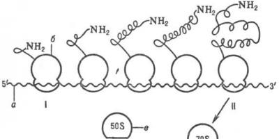
During protein synthesis, the ribosome moves along the thread-like mRNA molecule. The process is more efficient when not just one ribosome moves along the mRNA, but many ribosomes at the same time, resembling beads on a string in this case. Such chains of ribosomes are called polyribosomes or polysomes. On the endoplasmic reticulum, polysomes are found in the form of characteristic curls.
Ribosomal protein synthesis is a multistep process. The first stage (initiation) begins with the attachment of messenger RNA (mRNA) to the small ribosomal subunit, which is not associated with the large subunit. It is characteristic that a dissociated ribosome is required to begin the process. To the resulting so-called a large ribosomal subunit is attached to the initiation complex. Specialists participate in the initiation stage. initiation codon (see Genetic code), initiation transfer RNA (tRNA) and specific. proteins (so-called initiation factors). Having passed the initiation stage, the ribosome moves on to the sequence. reading the codons of mRNA in the direction from the 5" to the 3" end, which is accompanied by the synthesis of the polypeptide chain of the protein encoded by this mRNA (for more information about the mechanism of polypeptide synthesis, see article Translation). In this process, the ribosome functions as a cyclic molecule. car. The working cycle of the ribosome during elongation consists of three cycles: 1) codon-dependent binding of aminoacyl-tRNA (supplies amino acids to the ribosome), 2) transpeptidation - transfer of the C-terminus of the growing peptide to aminoacyl-tRNA, i.e. elongation of the protein chain under construction by one link, 3) translocation-movement of the matrix (mRNA) and peptidyl-tRNA relative to the ribosome and the transition of the ribosome to its original state, when it can perceive the trace. aminoacyl-tRNA. When the ribosome reaches the special stop codon of the mRNA, polypeptide synthesis stops. With the participation of specific proteins (so-called termination factors) synthesized. the polypeptide is released from the ribosome. After termination, the ribosome can repeat the entire cycle with another strand of mRNA or another coding sequence of the same strand.

Scheme of synthesis of a polypeptide chain by a polyribosome: I-beginning of synthesis, II-end of synthesis; a-mRNA, b-ribosome, b-large ribosomal subunit, g-small ribosomal subunit.
In cells with intense protein secretion and developed endoplasmic. reticulum means. part of the cytoplasmic ribosome is attached to its membrane on the surface facing the cytoplasm. These ribosomes synthesize polypeptides, which are directly transported across the membrane for further secretion. The synthesis of polypeptides for intracellular needs occurs mainly. on free (not membrane bound) ribosomes of the cytoplasm. In this case, translating ribosomes are not evenly dispersed in the cytoplasm, but are collected in groups. Such ribosome aggregates are structures where the mRNA is associated with many ribosomes that are in the process of translation; these structures are called polyribosome or polysome.
With intensive protein synthesis, the distance between ribosomes along the mRNA chain in a polyribosome may. extremely short, i.e. ribosomes are located almost close to each other. The ribosomes included in polyribosomes work independently and each of them synthesizes a complete polypeptide chain.
The ribosome is the very worker who puts the master plan into practice, producing the corresponding proteins based on DNA patterns.
Ribosomes are submicroscopic non-membrane organelles necessary for protein synthesis. They combine amino acids into a peptide chain to form new protein molecules. Biosynthesis is carried out using messenger RNA by translation.
Structural features
Ribosomes are located on the granular endoplasmic reticulum or float freely in the cytoplasm. They are attached to the endoplasmic reticulum with their large subunit and synthesize a protein that is transported outside the cell and used by the entire body. Cytoplasmic ribosomes mainly provide the internal needs of the cell.
The shape is spherical or oval, with a diameter of about 20 nm.
During the translation stage, several ribosomes can attach to the mRNA, forming a new structure - a polysome. They themselves are formed in the nucleolus, inside the nucleus.
There are 2 types of ribosomes:
- Small ones are found in prokaryotic cells, as well as in chloroplasts and the mitochondrial matrix. They are not associated with the membrane and are smaller in size (up to 15 nm in diameter).
- Large ones are found in eukaryotic cells, can reach a diameter of up to 23 nm, bind to the endoplasmic reticulum or are attached to the nuclear membrane.
 Structure diagram
 The structure of both types is identical. The ribosome consists of two subunits - large and small, which when combined resemble a mushroom. They are combined with the help of magnesium ions, maintaining a small gap between the contacting surfaces. With magnesium deficiency, the subunits move away, disaggregation occurs, and ribosomes can no longer perform their functions.
Chemical composition
Ribosomes consist of high-polymer ribosomal RNA and protein in a 1:1 ratio. They contain approximately 90% of all cellular RNA. The small and large subunits contain about four rRNA molecules, which look like threads gathered into a ball. The molecules are surrounded by proteins and together form a ribonucleoprotein.
Polyribosomes are a combination of messenger RNA and ribosomes that are strung on an mRNA strand. During the absence of synthesizing processes, ribosomes separate and exchange subunits. When mRNA arrives, they reassemble into polyribosomes.
The number of ribosomes can vary depending on the functional load on the cell. Tens of thousands are found in cells with high mitotic activity (plant meristem, stem cells).
Education in a cell
Ribosomal subunits are formed in the nucleolus. The template for the synthesis of ribosomal RNA is DNA. To fully mature, they go through several stages:
- Eosome is the first phase, in which only rRNA is synthesized on DNA in the nucleolus;
- neosome - a structure that includes not only rRNA, but also proteins, after a series of modifications it enters the cytoplasm;
- ribisome is a mature organelle consisting of two subunits.
Biosynthesis of proteins on ribosomes
Translation or synthesis of proteins on ribosomes from an mRNA matrix is the final stage in the transformation of genetic information in cells. During translation, information encoded in nucleic acids is transferred into protein molecules with a strict sequence of amino acids.
Translation is a very difficult stage (compared to replication and transcription). To carry out translation, all types of RNA, amino acids, and many enzymes are included in the process, which can correct each other’s errors. The most important participants in translation are ribosomes.
After transcription, the newly formed mRNA molecule leaves the nucleus into the cytoplasm. Here, after several transformations, it connects with the ribosome. In this case, amino acids are activated after interaction with the energy substrate - the ATP molecule.
Amino acids and mRNA have different chemical compositions and cannot interact with each other without outside participation. To overcome this incompatibility, transfer RNA exists. Under the action of enzymes, amino acids are combined with tRNA. In this form, they are transferred to the ribosome and tRNA, with a certain amino acid, is attached to the mRNA in the intended place. Next, ribosomal enzymes form a peptide bond between the attached amino acid and the polypeptide being built. The ribosome then moves along the messenger RNA chain, leaving a site for the attachment of the next amino acid.
The polypeptide grows until the ribosome encounters a “stop codon”, which signals the end of synthesis. To release the newly synthesized peptide from the ribosome, termination factors are activated, finally completing the biosynthesis. A water molecule is attached to the last amino acid, and the ribosome splits into two subunits.
As the ribosome moves further along the mRNA, it releases the initial section of the chain. A ribosome can again join it, which will begin a new synthesis. Thus, using one template for biosynthesis, ribosomes simultaneously create many copies of the protein.
The role of ribosomes in the body
- Ribosomes synthesize proteins for the cell's own needs and beyond. Thus, plasma blood clotting factors are formed in the liver, plasma cells produce gamma globulins.
- Reading encoded information from RNA, combining amino acids in a programmed order to form new protein molecules.
- Catalytic function – formation of peptide bonds, hydrolysis of GTP.
- Ribosomes perform their functions in the cell more actively in the form of polyribosomes. These complexes are capable of simultaneously synthesizing several protein molecules.
The ribosome, whose functions will be discussed in this article, is a particle that is located directly inside the cell. The main function of this particle is protein biosynthesis. The main one, but not the only one.
If we talk about the contribution of Russian scientists to the study of the ribosome, it is worth highlighting the work of the biochemist Academician A.S. Spirin.
Appearance of the ribosome and its other features
If you carefully examine the cell in electron micrographs, you can see small particles located in the cytoplasm. These particles are ribosomes.
The name "ribosome" consists of two parts. The first comes from “ribonucleic acid”, and the second is translated from Greek “soma” - body.
The size of ribonucleic particles in a cell ranges from 15-20 nm, and their quantity completely depends on the process of protein biosynthesis, namely its intensity. As a rule, there can be about 5,000 ribosomes, in some cases - up to 90,000. If we talk about the mass of this number of particles, it can sometimes reach a quarter of the mass of the cell itself.
The shape of the ribosome is more reminiscent of a sphere, but this fact cannot be stated unambiguously. But the function of ribosomes in a cell is associated with protein biosynthesis, and this is a confirmed fact.
By their chemical nature, these particles belong to nucleoproteins (a combination of nucleic acids with protein), which consist of ribonucleic acid.
Prokaryotic type
There are two types of ribosomes, the structure and functions of which are slightly different from each other.
The first type is characteristic of bacterial cells and green algae, that is, prokaryotic organisms. Its name is 70S ribosome, it performs the same functions. The number in the name means the sedimentation coefficient (a value that determines the size and shape of macromolecules, as well as the rate of sedimentation of a certain microparticle, in this case a ribosome, in a sufficiently strong gravitational field). For this type it is 70 Svedberg units. These ribosomes consist of two unequal particles: 30S and 50S. The first component contains one protein molecule, the second contains two RNA molecules. The main function performed by the protein molecules that make up the ribosome is structural.

Eukaryotic type
The second type of ribosomes was discovered in eukaryotic cells (plant or animal organisms whose cells have a clearly defined nucleus). The name of this subparticle is 80S. Ribosomes, whose functions are to synthesize proteins of this class, consist of equal parts of RNA and protein. But they also have the same two unequal subunits (60S and 40S).
Ribosomes: structure and functions
The ribosome consists of two unequal subunits.
The large subparticle, in turn, consists of:
- one molecule of ribosomal RNA, which is highly polymeric;
- one RNA molecule, which is low-polymer;
- a certain number of protein molecules, usually about three dozen.
As for the smaller subparticle, it’s a little simpler. It includes:
- high polymer RNA molecule;
- several dozen protein molecules, usually about 40 (the molecules are varied in structure and shape).

A high-polymer RNA molecule is necessary in order to combine all the proteins present into one integral ribonucleoprotein component of the cell.
In the process of performing its main function, that is, during protein synthesis, the ribosome also performs a number of additional ones:
- Ligament, as well as retention of all components of the so-called protein synthesizing system. It is customary to call this function informational or matrix. The ribosome distributes these functions between its two subparticles, each of which performs its own specific task in this process.
- Ribosomes perform a catalytic function, which consists in the formation of a special peptide bond (amide bond, which occurs both during the formation of proteins and during the formation of peptides). This also includes the hydrolysis of GTP (the substrate for RNA synthesis). The large subunit of the ribosome is responsible for performing this function. It is in it that there are special areas in which the process of peptide bond synthesis occurs, as well as the center necessary for the hydrolysis of GTP. In addition, it is the large subunit of the ribosome that, during protein biosynthesis, holds on itself a chain that gradually grows.
- The ribosome performs the functions of mechanical movement of substrates, which include mRNA and tRNA. In other words, they are responsible for translocation.

As a conclusion
Literally each of the ribosomal subunits, both large and small, can exhibit to some extent those functions that are directly associated with it, separately from its “neighbor”. However, only the complete ribosome can perform the translocation function.
We can safely say that there is a clear division of functions between the ribosomal particles. A small part is responsible for performing the reception, as well as deciphering genetic information. But the large particle is directly involved in transliteration.
In a bacterial cell, ribosomes make up up to 30% of its dry mass: there are approximately 10 4 ribosomes per bacterial cell. In eukaryotic cells (cells of all organisms, with the exception of bacteria and blue-green algae) refers. the content of ribosomes is smaller, and their number varies greatly depending on the protein-synthesizing activity of the corresponding tissue or individual cell.
In eukaryotic in a cell, all cytoplasmic ribosomes (both membrane-bound and free) are formed in the nucleolus; they are considered to be inactive there. Eukaryotic. the cell also has special ribosomes in mitochondria (in animals and plants) and chloroplasts (in plants). The ribosomes of these organelles differ from the cytoplasmic ones in size and certain functions. Holy you. They are formed directly in these organelles.
There are two main ones. type of ribosomes. Prokaryotic everyone. organisms (bacteria and blue-green algae) are characterized by the so-called. 70S ribosomes, characterized by coefficient. (constant) sedimentation approx. 70 Svedberg units, or 70S (based on the sedimentation coefficient, other types of ribosomes are also distinguished, as well as subparticles and biopolymers that make up ribosomes). They say m is 2.5 · 10 6, linear dimensions 20-25 nm. According to chemistry composition is ribonucleoproteins; they consist only of rRNA and protein (the ratio of these components is 2:1). Ribosomal RNA is present in ribosomes. arr. in the form of Mg-salt (apparently, partially also in the form of Ca-salt); magnesium in ribosomes up to 2% of dry weight. In addition, in different quantity (up to 2.5%) amine cations such as spermine H 2 N(CH 2) 3 NH(CH 2) 4 NH(CH 2) 3 NH 2 and spermidine H 2 N(CH 2) 3 NH may also be present (CH 2) 4 NH 2 etc.
Apparently, rRNA determines the basic structural and functional. The properties of ribosomes, in particular, ensure the integrity of ribosomal subunits, determine their shape and a number of structural features. Specific spaces. The rRNA structure determines the localization of all ribosomal proteins and plays a leading role in the organization of functions. ribosome centers.
Ribosomal protein synthesis is a multistep process. The first stage (initiation) begins with the attachment of messenger RNA (mRNA) to the small ribosomal subunit, which is not associated with the large subunit. It is characteristic that a dissociated ribosome is required to begin the process. To the resulting so-called a large ribosomal subunit is attached to the initiation complex. Specialists participate in the initiation stage. initiation codon (see Genetic code), initiation transfer RNA (tRNA) and specific. proteins (so-called initiation factors). Having passed the initiation stage, the ribosome moves on to the sequence. reading the codons of mRNA in the direction from the 5" to the 3" end, which is accompanied by the synthesis of the polypeptide chain of the protein encoded by this mRNA (for more information about the mechanism of polypeptide synthesis, see article Translation). In this process, the ribosome functions as a cyclic molecule. car. The working cycle of the ribosome during elongation consists of three cycles: 1) codon-dependent binding of aminoacyl-tRNA (supplies amino acids to the ribosome), 2) transpeptidation - transfer of the C-terminus of the growing peptide to aminoacyl-tRNA, i.e. elongation of the protein chain under construction by one link, 3) translocation-movement of the matrix (mRNA) and peptidyl-tRNA relative to the ribosome and the transition of the ribosome to its original state, when it can perceive the trace. aminoacyl-tRNA. When the ribosome reaches the special