Whatever you say, the microscope is one of the most important tools of scientists, one of their main weapons in understanding the world around us. How did the first microscope appear, what is the history of the microscope from the Middle Ages to the present day, what is the structure of the microscope and the rules for working with it, you will find answers to all these questions in our article. So let's get started.
The history of the microscope
Although the first magnifying lenses, on the basis of which the light microscope actually works, were found by archaeologists during the excavations of ancient Babylon, nevertheless, the first microscopes appeared in the Middle Ages. Interestingly, there is no agreement among historians as to who first invented the microscope. Among the candidates for this venerable role are such famous scientists and inventors as Galileo Galilei, Christian Huygens, Robert Hooke and Anthony van Leeuwenhoek.
It is also worth mentioning the Italian doctor G. Frakostoro, who, back in 1538, was the first to suggest combining several lenses in order to obtain a greater magnifying effect. This was not yet the creation of a microscope, but it became the forerunner of its occurrence.
And in 1590, a certain Hans Jasen, a Dutch master of eyeglasses, said that his son, Zakhary Yasen, invented the first microscope, for the people of the Middle Ages, such an invention was akin to a small miracle. However, a number of historians doubt whether Zachary Yasen is the true inventor of the microscope. The fact is that there are a lot of dark spots in his biography, including spots on his reputation, as contemporaries accused Zakharia of counterfeiting and stealing someone else's intellectual property. Be that as it may, but we, unfortunately, cannot find out for sure whether Zakhary Yasen was the inventor of the microscope or not.
But the reputation of Galileo Galilei in this regard is impeccable. We know this person, first of all, as a great astronomer, a scientist who was persecuted by the Catholic Church for his belief that the Earth revolves around, and not vice versa. Among the important inventions of Galileo is the first telescope, with the help of which the scientist penetrated the cosmic spheres with his gaze. But the scope of his interests was not limited to stars and planets, because a microscope is essentially the same telescope, but only the other way around. And if with the help of magnifying lenses you can observe distant planets, then why not turn their power in another direction - to study what is under our noses. “Why not,” Galileo probably thought, and now, in 1609, he was already presenting to the general public at the Accademia dei Licei his first compound microscope, which consisted of convex and concave magnifying lenses.

Vintage microscopes.
Later, 10 years later, the Dutch inventor Cornelius Drebbel improved Galileo's microscope by adding another convex lens to it. But the real revolution in the development of microscopes was made by Christian Huygens, a Dutch physicist, mechanic and astronomer. So he was the first to create a microscope with a two-lens system of eyepieces, which were regulated achromatically. It is worth noting that Huygens eyepieces are used to this day.
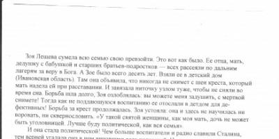
But the famous English inventor and scientist Robert Hooke entered the history of science forever, not only as the creator of his own original microscope, but also as a person who made a great scientific discovery with his help. It was he who first saw an organic cell through a microscope, and suggested that all living organisms consist of cells, these smallest units of living matter. Robert Hooke published the results of his observations in his fundamental work - Micrography.

Published in 1665 by the Royal Society of London, this book immediately became a scientific bestseller of those times and made a splash in the scientific community. No wonder, because it contained engravings depicting magnified under a microscope, lice, flies, plant cells. In fact, this work was an amazing description of the capabilities of the microscope.
An interesting fact: Robert Hooke took the term “cell” because plant cells bounded by walls reminded him of monastic cells.

This is what Robert Hooke's microscope looked like, image from Micrographia.
And the last outstanding scientist who contributed to the development of microscopes was the Dutchman Anthony van Leeuwenhoek. Inspired by Robert Hooke's Micrography, Leeuwenhoek created his own microscope. Leeuwenhoek's microscope, although it had only one lens, was extremely powerful, thus the level of detail and magnification of his microscope was the best at the time. Observing wildlife through a microscope, Leeuwenhoek made many important scientific discoveries in biology: he was the first to see erythrocytes, described bacteria, yeast, sketched spermatozoa and the structure of the eyes of insects, discovered and described many of their forms. Leeuwenhoek's work gave a huge impetus to the development of biology, and helped to attract the attention of biologists to the microscope, making it an integral part of biological research, even to this day. Such, in general terms, is the history of the discovery of the microscope.
Types of microscopes
Further, with the development of science and technology, more and more advanced light microscopes began to appear, the first light microscope, working on the basis of magnifying lenses, was replaced by an electronic microscope, and then a laser microscope, an X-ray microscope, giving many times better magnifying effect and detail. How do these microscopes work? More on this later.
Electron microscope
The history of the development of the electron microscope began in 1931, when a certain R. Rudenberg received a patent for the first transmission electron microscope. Then, in the 40s of the last century, scanning electron microscopes appeared, which reached their technical perfection already in the 60s of the last century. They formed an image of the object due to the successive movement of the electron probe of small cross section over the object.
How does an electron microscope work? Its work is based on a directed beam of electrons, accelerated in an electric field and displaying an image on special magnetic lenses, this electron beam is much smaller than the wavelength of visible light. All this makes it possible to increase the power of an electron microscope and its resolution by 1000-10,000 times compared to a traditional light microscope. This is the main advantage of the electron microscope.

This is what a modern electron microscope looks like.
laser microscope
The laser microscope is an improved version of the electron microscope; its operation is based on a laser beam, which allows the scientist's gaze to observe living tissues at an even greater depth.
X-ray microscope
X-ray microscopes are used to examine very small objects with dimensions comparable to those of an X-ray wave. Their work is based electromagnetic radiation with a wavelength from 0.01 to 1 nanometer.
Microscope device
The design of a microscope depends on its type, of course, an electron microscope will differ in its device from a light optical microscope or from an X-ray microscope. In our article, we will consider the structure of a conventional modern optical microscope, which is the most popular among both amateurs and professionals, since they can be used to solve many simple research problems.
So, first of all, in a microscope, one can distinguish the optical and mechanical parts. The optical part includes:
- The eyepiece is that part of the microscope that is directly connected to the eyes of the observer. In the very first microscopes, it consisted of a single lens; the design of the eyepiece in modern microscopes, of course, is somewhat more complicated.
- The lens is practically the most important part of the microscope, since it is the lens that provides the main magnification.
- Illuminator - responsible for the flow of light on the object under study.
- Aperture - regulates the strength of the light flux entering the object under study.
The mechanical part of the microscope consists of such important parts as:
- A tube is a tube that contains an eyepiece. The tube must be strong and not deform, otherwise the optical properties of the microscope will suffer.
- The base, it ensures the stability of the microscope during operation. It is on it that the tube, condenser holder, focusing knobs and other details of the microscope are attached.
- Turret - used for quick change of lenses, not available in cheap models of microscopes.
- The object table is the place on which the examined object or objects are placed.

And here the picture shows a more detailed structure of the microscope.
Rules for working with a microscope
- It is necessary to work with a microscope sitting;
- Before use, the microscope must be checked and dusted with a soft cloth;
- Set the microscope in front of you a little to the left;
- It is worth starting work with a small increase;
- Set the illumination in the field of view of the microscope using an electric illuminator or a mirror. Looking into the eyepiece with one eye and using a mirror with a concave side, direct the light from the window into the lens, and then illuminate the field of view as evenly and as much as possible. If the microscope is equipped with an illuminator, then connect the microscope to a power source, turn on the lamp and set the required brightness of combustion;
- Place the micropreparation on the stage so that the object under study is under the lens. Looking from the side, lower the lens with a macro screw until the distance between the lower lens of the objective and the micropreparation is 4-5 mm;
- Move the preparation by hand, find Right place, place it in the center of the field of view of the microscope;
- To study an object at high magnification, first place the selected area in the center of the microscope's field of view at low magnification. Then change the lens to 40 x by turning the revolver so that it is in its working position. Use a micrometer screw to achieve a good image of the object. On the box of the micrometer mechanism there are two dashes, and on the micrometer screw there is a dot, which must always be between the dashes. If it goes beyond their limits, it must be returned to its normal position. If this rule is not observed, the micrometer screw may stop working;
- Upon completion of work with a high magnification, set a low magnification, raise the lens, remove the preparation from the working table, wipe all parts of the microscope with a clean cloth, cover it with a plastic bag and put it in a cabinet.

When writing the article, I tried to make it as interesting, useful and of high quality as possible. I will be grateful for any feedback and constructive criticism in the form of comments to the article. You can also write your wish / question / suggestion to my mail [email protected] or on Facebook, with respect, the author.
MICROSCOPE
REPORT on Biology of a 6th grade student
For a long time, a person lived surrounded by invisible creatures, used their waste products (for example, when baking bread from sour dough, making wine and vinegar), suffered when these creatures caused illnesses or spoiled food supplies, but did not suspect their presence . I didn't suspect because I didn't see it, and I didn't see it because the sizes of these micro creatures were much lower than the limit of visibility that the human eye is capable of. It is known that a person with normal vision at the optimal distance (25–30 cm) can distinguish an object 0.07–0.08 mm in size in the form of a point. Smaller objects cannot be seen. This is determined by the structural features of his organ of vision.
Approximately at the same time when the exploration of space with the help of telescopes began, the first attempts were made to reveal, with the help of lenses, the secrets of the microworld. Thus, during archaeological excavations in Ancient Babylon biconvex lenses were found - the simplest optical devices. The lenses were made from polished mountain crystal. It can be considered that with their invention man took the first step on the way to the microworld.
The simplest way to enlarge the image of a small object is to observe it with a magnifying glass. A magnifying glass is a converging lens with a small focal length (usually no more than 10 cm) inserted into the handle.
telescope maker Galileo V 1610
In 1993, he discovered that, when wide apart, his spotting scope made it possible to greatly enlarge small objects. It can be considered the inventor of the microscope consisting of positive and negative lenses.
A more advanced tool for observing microscopic objects is simple microscope. When these devices appeared, it is not known exactly. At the very beginning of the 17th century, several such microscopes were made by a spectacle craftsman Zacharias Jansen from Middelburg.
In the essay A. Kircher, released in 1646 year, contains a description the simplest microscope named by him "flea glass". It consisted of a magnifying glass embedded in a copper base, on which an object table was fixed, which served to place the object in question; at the bottom there was a flat or concave mirror, reflecting the sun's rays onto an object and thus illuminating it from below. The magnifying glass was moved by means of a screw to the object table until the image became distinct and clear.
First great discoveries were just made using a simple microscope. In the middle of the 17th century, brilliant success was achieved by the Dutch naturalist Anthony Van Leeuwenhoek. For many years, Leeuwenhoek perfected himself in the manufacture of tiny (sometimes less than 1 mm in diameter) biconvex lenses, which he made from a small glass ball, which in turn was obtained by melting a glass rod in a flame. Then this glass ball was ground on a primitive grinding machine. During his life, Leeuwenhoek made at least 400 such microscopes. One of them, kept in the University Museum in Utrecht, gives more than 300x magnification, which was a huge success for the 17th century.
At the beginning of the 17th century, there were compound microscopes composed of two lenses. The inventor of such a complex microscope is not exactly known, but many facts indicate that he was a Dutchman. Cornelius Drebel, who lived in London and was in the service of the English King James I. In the compound microscope, there was two glasses: one - the lens - facing the object, the other - the eyepiece - facing the eye of the observer. In the first microscopes, a biconvex glass served as an objective, which gave a real, enlarged, but inverse image. This image was examined with the help of an eyepiece, which thus played the role of a magnifying glass, but only this magnifying glass served to magnify not the object itself, but its image.
IN 1663 microscope Drebel was improved English physicist Robert Hooke, who introduced a third lens into it, called the collective. This type of microscope gained great popularity, and most of the microscopes of the late 17th - first half of the 8th century were built according to its scheme.
Microscope device
A microscope is an optical instrument designed to study magnified images of micro-objects that are invisible to the naked eye.
The main parts of a light microscope (Fig. 1) are an objective and an eyepiece enclosed in a cylindrical body - a tube. Most models designed for biological research come with three lenses with different focal lengths and a rotating mechanism designed for quick change - a turret, often called a turret. The tube is located on the top of a massive stand, including the tube holder. Slightly below the objective (or turret with multiple objectives) is an object stage, on which slides with test samples are placed. Sharpness is adjusted using a coarse and fine adjustment screw, which allows you to change the position of the stage relative to the objective.
In order for the sample under study to have sufficient brightness for comfortable observation, the microscopes are equipped with two more optical units (Fig. 2) - an illuminator and a condenser. The illuminator creates a stream of light that illuminates the test preparation. In classical light microscopes, the design of the illuminator (built-in or external) involves a low-voltage lamp with a thick filament, a converging lens, and a diaphragm that changes the diameter of the light spot on the sample. The condenser, which is a converging lens, is designed to focus the illuminator beams on the sample. The condenser also has an iris diaphragm (field and aperture), which controls the intensity of illumination.
When working with light-transmitting objects (liquids, thin sections of plants, etc.), they are illuminated by transmitted light - the illuminator and condenser are located under the object stage. Opaque samples should be illuminated from the front. To do this, the illuminator is placed above the object stage, and its beams are directed to the object through the lens using a translucent mirror.
The illuminator may be passive, active (lamp), or both. The simplest microscopes do not have lamps to illuminate samples. Under the table they have a double-sided mirror, in which one side is flat and the other is concave. In daylight, if the microscope is near a window, you can get pretty good illumination using a concave mirror. If the microscope is in a dark room, a flat mirror and an external illuminator are used for illumination.
The magnification of a microscope is equal to the product of the magnification of the objective and the eyepiece. With an eyepiece magnification of 10 and an objective magnification of 40, the total magnification factor is 400. Usually, objectives with a magnification of 4 to 100 are included in a research microscope kit. A typical microscope objective kit for amateur and educational research (x4, x10 and x40), provides increase from 40 to 400.
Resolution is another important characteristic of a microscope, which determines its quality and the clarity of the image it forms. The higher the resolution, the more fine details can be seen at high magnification. In connection with resolution, one speaks of "useful" and "useless" magnification. “Useful” is the maximum magnification at which the maximum image detail is provided. Further magnification (“useless”) is not supported by the resolution of the microscope and does not reveal new details, but it can adversely affect the clarity and contrast of the image. Thus, the limit of useful magnification of a light microscope is not limited overall coefficient magnification of the lens and eyepiece - it can be made as large as desired if desired - but the quality of the optical components of the microscope, that is, the resolution.
The microscope includes three main functional parts:
1. Lighting part
Designed to create a light flux that allows you to illuminate the object in such a way that the subsequent parts of the microscope perform their functions with the utmost accuracy. The illuminating part of a transmitted light microscope is located behind the object under the objective in direct microscopes and in front of the object above the objective in inverted ones.
The lighting part includes a light source (a lamp and an electric power supply) and an optical-mechanical system (collector, condenser, field and aperture adjustable / iris diaphragms).
2. Playback part
Designed to reproduce an object in the image plane with the image quality and magnification required for research (i.e., to build such an image that reproduces the object as accurately as possible and in all details with the resolution, magnification, contrast and color reproduction corresponding to the microscope optics).
The reproducing part provides the first stage of magnification and is located after the object to the image plane of the microscope. The reproducing part includes a lens and an intermediate optical system.
Modern microscopes of the latest generation are based on optical systems of lenses corrected for infinity.
This additionally requires the use of so-called tube systems, which “collect” parallel beams of light coming out of the objective in the image plane of the microscope.
3. Visualizing part
Designed to obtain a real image of an object on the retina, film or plate, on the screen of a television or computer monitor with additional magnification (the second stage of magnification).
The imaging part is located between the image plane of the lens and the eyes of the observer (camera, camera).
The imaging part includes a monocular, binocular or trinocular visual attachment with an observation system (eyepieces that work like a magnifying glass).
In addition, this part includes systems of additional magnification (systems of a wholesaler / change of magnification); projection nozzles, including discussion nozzles for two or more observers; drawing devices; image analysis and documentation systems with appropriate matching elements (photo channel).
Histology how an independent science emerged in early XIX century. The prehistory of histology was the results of numerous macroscopic (visual) studies of the constituent parts of various animal and plant organisms. Of decisive importance for the development of histology as a science of the structure of tissues was the invention of the microscope, the first samples of which were created at the beginning of the 17th century (G. and Z. Jansen, G. Galilei, and others). One of the earliest scientific studies using a microscope of his own design was carried out by the English scientist Robert Hooke (1635-1703). He studied the microscopic structure of many objects. R. Hooke described all the objects studied in the book "Micrography or some physiological descriptions of the smallest bodies made with the help of magnifying glasses ...", published in 1665. From his observations, R. Hooke concluded that bubble-shaped cells, or cells, are widespread, in plant objects and first proposed the term "cell".
In 1671, the English scientist N. Grew (1641-1712) in his book " plant anatomy" wrote about cellular structure as a general principle of organization of plant organisms. N. Gru first introduced the term "fabric" to denote plant mass, since the latter resembled clothing fabrics in its microscopic design. In the same year, the Italian J. Malpighi (1628-1694) gave a systematic and detailed description cellular (cellular) structure of various plants. In the future, facts gradually accumulated, indicating that not only plant, but also animal organisms consist of cells. In the second half of the 17th century, A. Leeuwenhoek (1632-1723) discovered the world of microscopic animals and for the first time described red blood cells and male sex cells.
Throughout the 18th century there was a gradual accumulation of facts about the cellular structure of plants and animals. Cells of animal tissues were studied and described in detail by the Czech scientist Jan Purkynia (1787-1869) and his students at the beginning of the 19th century.
Of great importance for the development of knowledge about microscopic structure of organisms has further improved microscopes. In the 18th century, microscopes were being produced already in in large numbers. They were first brought to Russia from Holland by Peter I. Later, a workshop for the manufacture of microscopes was organized at the Academy of Sciences in St. Petersburg. M.V. did a lot for the development of microscopy in Russia. Lomonosov, who proposed a number of technical improvements in the design of the microscope and its optical system. The second half of the 19th century is notable for the rapid improvement of microscopic technology. New designs of microscopes were created, and thanks to the invention of immersion lenses (water immersion began to be used from 1850, oil immersion - from 1878), the resolution of optical instruments increased tenfold. In parallel with the improvement of the microscope, the technique of preparing microscopic preparations also developed.
If earlier objects examined under a microscope immediately after their isolation from plants or animals without any preliminary preparation, now they began to resort to various methods of processing them, which made it possible to preserve the structure of biological objects. Various methods of material fixation have been proposed. Chromic, picric, osmic, acetic and other acids, as well as their mixtures, have been used as fixing agents. A simple and in many cases indispensable fixative - formalin - was first used to fix biological objects in 1893.
Manufacturing of drugs, suitable for examination in transmitted light, became possible after the development of methods for pouring pieces into dense media, which made it easier to obtain thin sections. The invention of special structures for cutting - microtomes - in the laboratory of J. Purkins significantly improved the technique for making histological preparations. In Russia, the first microtome was constructed by the Kiev histologist P.I. Peremezhko. To enhance the contrast of the structures, the sections began to be stained with various dyes. Carmine was the first histological dye that stained cell nuclei and was widely used (beginning in 1858). Another nuclear dye - hematoxylin - has been used since 1865, but for a long time its properties were not fully evaluated. By the second half of the 19th century, aniline dyes were already used, a method was developed for impregnating tissues with silver nitrate (K. Golgi, 1873) and staining of nervous tissue with methylene blue (A.S. Dogel, A.E. Smirnov, 1887).
Thanks to fixation biological material and obtaining the thinnest colored sections from it, researchers of the late 19th century had the opportunity to penetrate much deeper into the secrets of the structure of tissues and cells, on the basis of which a number of the greatest discoveries were made. So, in 1833, R. Brown discovered a permanent component of the cell - the nucleus. In 1861, M. Schultze approved the view of the cell as "a lump of protoplasm with a nucleus lying inside it." Main constituent parts cells began to count the nucleus and cytoplasm. In the 70s of the XIX century, a group of researchers simultaneously and independently discovered an indirect method of cell division - karyokinesis, or mitosis. In the works of I.D. Chistyakov (1874), O. Buchli (1875), E. Strasburger (1875), W. Meisel (1875), P.I. Peremezhko (1878), V. Schleicher (1878), V. Flemming (1879) and others described and illustrated all stages of indirect cell division. This discovery had great importance to develop knowledge about the cell. It also served as the basis for a deeper study of such an important biological process as fertilization. The study of mitosis and fertilization attracted particular attention of researchers to the cell nucleus and elucidation of its significance in the process of transferring hereditary properties. In 1884, O. Gertwig and E. Strasburger independently put forward the hypothesis that chromatin is the material carrier of heredity.
The object of close attention of scientists is chromosomes. Along with the study of the cell nucleus, the cytoplasm was also subjected to a thorough analysis.
Advances in microscopic technology have led to opening of organelles in the cytoplasm- its constant and highly differentiated elements, having a certain structure and performing vital functions for the cell. In 1875-76. the German biologist O. Hertwig and the Belgian scientist Van Beneden discovered the cell center, or centrosome; and in 1898 by the Italian scientist K. Golgi - the intracellular reticular apparatus (Golgi complex). In 1897, K. Benda - in animal cells, and in 1904 - F. Mewes - in plant cells described chondriosomes, which later became known as mitochondria.
Thus, by the end of the 19th century, on the basis of the successful development of microscopic technology and analysis of data on the microscopic structure of the cell, colossal factual material was accumulated, which made it possible to identify a number of important patterns in the structure and development of cells and tissues. At this time, the doctrine of the cell stood out in an independent biological science - cytology.
This is the science of life. At present, it represents the totality of the sciences of living nature.
Biology studies all manifestations of life: structure, functions, development and origin living organisms, their relationship in natural communities with the environment and with other living organisms.
Since man began to realize his difference from the animal world, he began to study the world around him.
At first, his life depended on it. Primitive people needed to know which living organisms can be eaten, used as medicines, for making clothes and dwellings, and which of them are poisonous or dangerous.
With the development of civilization, a person could afford such a luxury as doing science for educational purposes.
Research the cultures of the ancient peoples showed that they had extensive knowledge about plants and animals and widely applied them in everyday life.
Modern biology - complex the science, which is characterized by the interpenetration of ideas and methods of various biological disciplines, as well as other sciences - primarily physics, chemistry and mathematics.
The main directions of development of modern biology. Currently, three directions in biology can be conditionally distinguished.
First, it is classical biology. It is represented by natural scientists who study the diversity of living nature. They objectively observe and analyze everything that happens in wildlife, study living organisms and classify them. It is wrong to think that in classical biology all discoveries have already been made.
In the second half of the XX century. not only many new species have been described, but also large taxa have been discovered, up to kingdoms (Pogonophores) and even superkingdoms (Archaebacteria, or Archaea). These discoveries forced scientists to take a fresh look at the entire development history living nature, For true natural scientists, nature is a value in itself. Every corner of our planet is unique for them. That is why they are always among those who acutely feel the danger to the nature around us and actively advocate for it.
The second direction is evolutionary biology.
In the 19th century author of the theory natural selection Charles Darwin started out as an ordinary naturalist: he collected, observed, described, traveled, revealing the secrets of wildlife. However, the main result of his work that made him a famous scientist was the theory explaining organic diversity.

Currently, the study of the evolution of living organisms is actively continuing. The synthesis of genetics and evolutionary theory led to the creation of the so-called synthetic theory of evolution. But even now there are still many unresolved questions that evolutionary scientists are looking for answers to.

Created at the beginning of the 20th century. our outstanding biologist Alexander Ivanovich Oparin, the first scientific theory The origin of life was purely theoretical. Currently being actively experimental studies of this problem and thanks to the use of advanced physical and chemical methods have already been made important discoveries and we can expect new interesting results.
New discoveries made it possible to supplement the theory of anthropogenesis. But the transition from the animal world to man still remains one of the biggest mysteries of biology.

The third direction is physical and chemical biology, which studies the structure of living objects using modern physical and chemical methods. This is a rapidly developing area of biology, important both in theoretical and practical terms. We can say with confidence that new discoveries await us in physical and chemical biology, which will allow us to solve many problems facing humanity.

The development of biology as a science. Modern biology is rooted in antiquity and is associated with the development of civilization in the Mediterranean countries. We know the names of many outstanding scientists who contributed to the development of biology. Let's name just a few of them.
Hippocrates (460 - c. 370 BC) gave the first regarding detailed description structure of man and animals, pointed to the role of the environment and heredity in the occurrence of diseases. He is considered the founder of medicine.

Aristotle (384-322 BC) divided the world into four kingdoms: the inanimate world of earth, water and air; plant world; the animal world and the human world. He described many animals, laid the foundation for taxonomy. The four biological treatises he wrote contained almost all the information about animals known by that time. The merits of Aristotle are so great that he is considered the founder of zoology.
Theophrastus (372-287 BC) studied plants. He described more than 500 plant species, gave information about the structure and reproduction of many of them, introduced many botanical terms. He is considered the founder of botany.

Gaius Pliny the Elder (23-79) collected information about living organisms known by that time and wrote 37 volumes of the Natural History Encyclopedia. Almost until the Middle Ages, this encyclopedia was the main source of knowledge about nature.
Claudius Galen made extensive use of dissections of mammals in his scientific research. He was the first to make a comparative anatomical description of man and monkey. Studied central and peripheral nervous system. Historians of science consider him the last great biologist of antiquity.
In the Middle Ages, religion was the dominant ideology. Like other sciences, biology during this period had not yet emerged as an independent field and existed in the general mainstream of religious and philosophical views. And although the accumulation of knowledge about living organisms continued, one can speak of biology as a science at that time only conditionally.
The Renaissance is a transitional period from the culture of the Middle Ages to the culture of modern times. The fundamental socio-economic transformations of that time were accompanied by new discoveries in science.
The most famous scientist of this era, Leonardo da Vinci (1452 - 1519), made a certain contribution to the development of biology.
He studied the flight of birds, described many plants, ways of connecting bones in the joints, the activity of the heart and the visual function of the eye, the similarity of human and animal bones.
In the second half of the XV century. natural sciences begin to develop rapidly. This was facilitated geographical discoveries, which made it possible to significantly expand information about animals and plants. The rapid accumulation of scientific knowledge about living organisms led to the division of biology into separate sciences.
In the XVI-XVII centuries. Botany and zoology began to develop rapidly.
The invention of the microscope (early 17th century) made it possible to study the microscopic structure of plants and animals. Microscopically small living organisms, bacteria and protozoa, invisible to the naked eye, were discovered.
A great contribution to the development of biology was made by Carl Linnaeus, who proposed a classification system for animals and plants,
Karl Maximovich Baer (1792-1876) in his works formulated the main provisions of the theory of homologous organs and the law of germinal similarity, which laid the scientific foundations of embryology.
In 1808, in his Philosophy of Zoology, Jean-Baptiste Lamarck raised the question of the causes and mechanisms of evolutionary transformations and outlined the first theory of evolution in time.
The cell theory played a huge role in the development of biology, which scientifically confirmed the unity of the living world and served as one of the prerequisites for the emergence of Charles Darwin's theory of evolution. The zoologist Theodor Ivann (1818-1882) and the botanist Matthias Jakob Schleiden (1804-1881) are considered the authors of the cell theory.
On the basis of numerous observations, Charles Darwin published in 1859 his main work “On the Origin of Species by Means of Natural Selection, or the Preservation of Favored Breeds in the Struggle for Life”, in which he formulated the main provisions of the theory of evolution, proposed the mechanisms of evolution and ways of evolutionary transformations of organisms.
In the 19th century Thanks to the work of Louis Pasteur (1822-1895), Robert Koch (1843-1910), Ilya Ilyich Mechnikov, microbiology took shape as an independent science.
The 20th century began with the rediscovery of Gregor Mendel's laws, which marked the beginning of the development of genetics as a science.
In the 40-50s of the XX century. in biology, the ideas and methods of physics, chemistry, mathematics, cybernetics and other sciences began to be widely used, and microorganisms were used as objects of study. As a result, biophysics, biochemistry, molecular biology, radiation biology, bionics, etc. emerged and rapidly developed as independent sciences. Space exploration contributed to the birth and development of space biology.
In the XX century. the direction of applied research - biotechnology. This trend will undoubtedly develop rapidly in the 21st century. You will learn more about this direction in the development of biology when studying the chapter "Fundamentals of Breeding and Biotechnology".
Currently, biological knowledge is used in all areas human activity: in industry and agriculture, medicine and energy.
Ecological research is extremely important. We finally began to realize that the delicate balance that exists on our small planet is easy to destroy. Mankind has faced a daunting task - the preservation of the biosphere in order to maintain the conditions for the existence and development of civilization. It is impossible to solve it without biological knowledge and special studies. Thus, at present, biology has become a real productive force and a rational scientific basis for the relationship between man and nature.
classical biology. Evolutionary biology. Physical and chemical biology.
1. What directions in the development of biology can you single out?
2. What great scientists of antiquity made a significant contribution to the development of biological knowledge?
3. Why in the Middle Ages it was possible to speak about biology as a science only conditionally?
4. Why modern biology considered a complex science?
5. What is the role of biology in modern society?
6. Prepare a message on one of the following topics:
7. The role of biology in modern society.
8. The role of biology in space research.
9. The role of biological research in modern medicine.
10. The role of outstanding biologists - our compatriots in the development of world biology.
How much the views of scientists on the diversity of living things have changed can be demonstrated by the example of the division of living organisms into kingdoms. Back in the 40s of the XX century, all living organisms were divided into two kingdoms: Plants and Animals. The plant kingdom also included bacteria and fungi. Later, a more detailed study of organisms led to the allocation of four kingdoms: Prokaryotes (Bacteria), Fungi, Plants and Animals. This system given in school biology.
In 1959, it was proposed to divide the world of living organisms into five kingdoms: Prokaryotes, Protists (Protozoa), Fungi, Plants and Animals.
This system is often given in biological (especially translated) literature.
Other systems have been developed and continue to be developed, including 20 or more kingdoms. For example, it is proposed to distinguish three superkingdoms: Prokaryotes, Archaea (Archaebacteria) and Eukaryotes. Each superkingdom includes several kingdoms.
Kamensky A. A. Biology grade 10-11
Submitted by readers from the website
Online library with students and books, outlines of lessons from Grade 10 Biology, books and textbooks according to the calendar plan planning Biology Grade 10
Lesson content lesson summary and support frame lesson presentation interactive technologies accelerating teaching methods Practice quizzes, testing online tasks and exercises homework workshops and trainings questions for class discussions Illustrations video and audio materials photos, pictures graphics, tables, schemes comics, parables, sayings, crossword puzzles, anecdotes, jokes, quotes Add-onsIt's hard to imagine today scientific activity man without a microscope. The microscope is widely used in most laboratories of medicine and biology, geology and materials science.
The results obtained using a microscope are necessary for making an accurate diagnosis and monitoring the course of treatment. With the use of a microscope, new drugs are developed and introduced, scientific discoveries are made.
Microscope- (from the Greek mikros - small and skopeo - I look), an optical device for obtaining an enlarged image of small objects and their details that are not visible to the naked eye.
The human eye is able to distinguish the details of an object that are at least 0.08 mm apart from each other. Using a light microscope, you can see the details, the distance between which is up to 0.2 microns. An electron microscope allows you to get a resolution of up to 0.1-0.01 nm.
The invention of the microscope, an instrument so important for all science, is primarily due to the influence of the development of optics. Some optical properties of curved surfaces were known even to Euclid (300 BC) and Ptolemy (127-151), but their magnifying power was not found practical application. In this regard, the first glasses were invented by Salvinio deli Arleati in Italy only in 1285. In the 16th century, Leonardo da Vinci and Maurolico showed that small objects are best studied with a magnifying glass.
 The first microscope was created only in 1595 by Z. Jansen. The invention consisted in the fact that Zacharius Jansen mounted two convex lenses inside one tube, thereby laying the foundation for the creation of complex microscopes. Focusing on the object under study was achieved by a retractable tube. The magnification of the microscope was from 3 to 10 times. And it was a real breakthrough in the field of microscopy! Each of his next microscope, he significantly improved.
During this period (XVI century) Danish, English and Italian research instruments gradually began to develop, laying the foundation for modern microscopy.
The rapid spread and improvement of microscopes began after Galileo (G. Galilei), improving the telescope he designed, began to use it as a kind of microscope (1609-1610), changing the distance between the objective and the eyepiece.
Later, in 1624, having achieved the manufacture of shorter focus lenses, Galileo significantly reduced the dimensions of his microscope.
In 1625, a member of the Roman Academy of the Vigilant ("Akudemia dei lincei") I. Faber proposed the term "microscope". The first successes associated with the use of a microscope in scientific biological research, were achieved by R. Hooke, who was the first to describe the plant cell (circa 1665). In his book "Micrographia" Hooke described the structure of the microscope.
 In 1681, the Royal Society of London in their meeting discussed in detail the peculiar situation. Dutchman Leeuwenhoek(A. van Leenwenhoek) described the amazing miracles that he discovered with his microscope in a drop of water, in an infusion of pepper, in the mud of a river, in the hollow of his own tooth. Leeuwenhoek, using a microscope, discovered and sketched the spermatozoa of various protozoa, details of the structure of bone tissue (1673-1677).
"With the greatest amazement, I saw in the drop a great many small animals moving briskly in all directions, like a pike in water. The smallest of these tiny animals is a thousand times smaller than the eye of an adult louse."
The best Leeuwenhoek magnifiers were magnified 270 times. With them, he saw for the first time the blood corpuscles, the movement of blood in the capillary vessels of the tail of the tadpole, the striation of the muscles. He opened infusoria. For the first time he plunged into the world of microscopic unicellular algae, where the border between animal and plant lies; where a moving animal, like a green plant, has chlorophyll and feeds by absorbing light; where the plant, still attached to the substrate, has lost chlorophyll and is ingesting bacteria. Finally, he even saw bacteria in great variety. But, of course, at that time there was still no remote possibility of understanding either the significance of bacteria for humans, or the meaning of the green substance - chlorophyll, or the boundary between plant and animal.
opened new world living beings, more varied and infinitely more original than the world we see.
In 1668, E. Divini, having attached a field lens to the eyepiece, created an eyepiece of the modern type. In 1673, Haveliy introduced a micrometer screw, and Hertel suggested placing a mirror under the microscope stage. Thus, the microscope began to be assembled from those main parts that are part of a modern biological microscope.
 In the middle of the 17th century newton discovered the complex composition of white light and decomposed it with a prism. Römer proved that light travels at a finite speed and measured it. Newton made the famous hypothesis - wrong, as you know - that light is a stream of flying particles of such extraordinary fineness and frequency that they penetrate through transparent bodies, like glass through the lens of the eye, and, hitting the retina with blows, produce physiological sensation Sveta. Huygens was the first to speak of the undulating nature of light and proved how naturally it explains both the laws of simple reflection and refraction, and the laws of double refraction in Icelandic spar. The thoughts of Huygens and Newton met in sharp contrast. Thus, in the XVII century. in a sharp dispute, the problem of the essence of light really arose.
Both the solution to the question of the essence of light and the improvement of the microscope moved forward slowly. The dispute between the ideas of Newton and Huygens continued for a century. The famous Euler joined the idea of the wave nature of light. But the issue was resolved only after more than a hundred years by Fresnel, a talented researcher, such as science knew.
What is the difference between the flow of propagating waves - the idea of Huygens - from the flow of rushing small particles - the idea of Newton? Two signs:
1. Having met, the waves can mutually annihilate if the hump of one lies on the valley of the other. Light + light combined together can produce darkness. This phenomenon interference, these are Newton's rings, misunderstood by Newton himself; this cannot be the case with particle flows. Two streams of particles are always a double stream, a double light.
2. The flow of particles passes through the hole directly, without diverging to the sides, and the flow of waves certainly diverges, dissipates. This diffraction.
Fresnel proved theoretically that the divergence in all directions is negligible if the wave is small, but nevertheless he discovered and measured this negligible diffraction, and determined the wavelength of light from its magnitude. Of the interference phenomena that are so well known to opticians who polish to "one color", to "two bands", he also measured the wavelength - this is half a micron (half a thousandth of a millimeter). And hence the wave theory and the exceptional subtlety and sharpness of penetration into the essence of living matter became undeniable. Since then, we all confirm and apply Fresnel's ideas in various modifications. But even without knowing these thoughts, one can improve the microscope.
So it was in the 18th century, although events developed very slowly. Now it is difficult even to imagine that Galileo's first tube, through which he observed the world of Jupiter, and Leeuwenhoek's microscope were simple non-achromatic lenses.
A huge obstacle to achromatization was the lack of a good flint. As you know, achromatization requires two glasses: crown and flint. The latter is glass, in which one of the main parts is heavy lead oxide, which has a disproportionately large dispersion.
 In 1824, Sallig's simple practical idea, reproduced by the French firm of Chevalier, gave a tremendous success to the microscope. The lens, which used to consist of a single lens, is divided into parts, it began to be made from many achromatic lenses. Thus, the number of parameters was multiplied, the possibility of correcting system errors was given, and for the first time it became possible to talk about real large magnifications - by 500 and even 1000 times. The boundary of ultimate vision has moved from two to one micron. Leeuwenhoek's microscope is left far behind.
In the 70s of the 19th century, the victorious march of microscopy moved forward. The one who said was Abbe(E. Abbe).
The following has been achieved:
First, the limiting resolution has moved from half a micron to one tenth of a micron.
Secondly, in the construction of the microscope, instead of rough empiricism, a high scientific character has been introduced.
Thirdly, finally, the limits of the possible with a microscope are shown, and these limits are conquered.
A headquarters of scientists, opticians and calculators working at the Zeiss firm was formed. Abbe's pupils presented the theory of the microscope and of optical instruments in general in major works. A system of measurements has been developed that determines the quality of a microscope.
When it turned out that the existing types of glass could not satisfy scientific requirements, new varieties were systematically created. Outside the secrets of Guinan's heirs - Para-Mantua (the heirs of Bontan) in Paris and Chance in Birmingham - glass melting methods were again created, and the matter of practical optics was developed to such an extent that one can say: Abbe almost won with the optical equipment of the army world war 1914-1918
Finally, calling to the aid of the foundations of the wave theory of light, Abbe clearly showed for the first time that each sharpness of the instrument has its own limit of possibility. The thinnest of all instruments is the wavelength. It is impossible to see objects less than half the wavelength, says Abbe's diffraction theory, and it is impossible to obtain images less than half the wavelength, i.e. less than 1/4 micron. Or with various tricks of immersion, when we use media in which the wavelength is shorter - up to 0.1 micron. The wave limits us. True, the limits are very small, but still these are limits for human activity.
 An optical physicist feels when an object a thousandth, ten thousandth, in some cases even one hundred thousandth of a wavelength is inserted in the path of a light wave. The wavelength itself is measured by physicists with an accuracy of one ten-millionth of its magnitude. Is it possible to think that opticians, who have joined their efforts with cytologists, will not master the hundredth wavelength that stands in their task? There are dozens of ways to get around the wavelength limit. You know one of these bypasses, the so-called ultramicroscopy method. If the microbes invisible in the microscope are far apart, then you can illuminate them from the side with a bright light. No matter how small they are, they will shine like a star against a dark background. Their form cannot be determined, one can only ascertain their presence, but this is often extremely important. This method is widely used in bacteriology.
The works of the English optician J. Sirks (1893) laid the foundation for interference microscopy. In 1903 R. Zsigmondy and N. Siedentopf created an ultramicroscope, in 1911 M. Sagnac described the first two-beam interference microscope, in 1935 F. Zernicke proposed use the phase contrast method to observe transparent, weakly light-scattering objects in microscopes. In the middle of the XX century. the electron microscope was invented, in 1953 the Finnish physiologist A. Wilska invented the anoptral microscope.
M.V. Lomonosov, I.P. Kulibin, L.I. Mandelstam, D.S. Rozhdestvensky, A.A. Lebedev, S.I. Vavilov, V.P. Linnik, D.D. Maksutov and others.
Literature:
D.S. Christmas Selected writings. M.-L., "Science", 1964.
Rozhdestvensky D.S. On the question of the image of transparent objects in a microscope. - Tr. GOI, 1940, v. 14
Sobol S.L. History of the microscope and microscopic research in Russia in the 18th century. 1949.
Clay R.S., Court T.H. The history of the microscope. L., 1932; Bradbury S. The evolution of the microscope. Oxford, 1967.