Length and distance converter Mass converter Converter of volume measures of bulk products and food products Area converter Converter of volume and units of measurement in culinary recipes Temperature converter Converter of pressure, mechanical stress, Young's modulus Converter of energy and work Converter of power Converter of force Converter of time Linear speed converter Flat angle Converter thermal efficiency and fuel efficiency Converter of numbers in various number systems Converter of units of measurement of quantity of information Exchange rates Sizes of women's clothing and shoes Sizes men's clothing and shoes Angular velocity and rotational speed converter Acceleration converter Angular acceleration converter Density converter Specific volume converter Moment of inertia converter Moment of force converter Torque converter Specific heat of combustion converter (by mass) Energy density and specific heat of combustion converter of fuel (by volume) Temperature difference converter Thermal expansion coefficient converter Thermal resistance converter Thermal conductivity converter Specific heat capacity converter Energy exposure and thermal radiation power converter Heat flux density converter Heat transfer coefficient converter Volume flow rate converter Mass flow rate converter Molar flow rate converter Mass flow density converter Molar concentration converter Mass concentration in solution converter Dynamic flow rate converter (absolute) viscosity Kinematic viscosity converter Surface tension converter Vapor permeability converter Water vapor flux density converter Sound level converter Microphone sensitivity converter Sound pressure level (SPL) converter Sound pressure level converter with selectable reference pressure Brightness converter Luminous intensity converter Illumination converter Computer resolution converter graph Frequency and wavelength converter Optical power in diopters and focal length Optical power in diopters and lens magnification (×) Converter electric charge Linear Charge Density Converter Surface Charge Density Converter Volume Charge Density Converter Converter electric current Linear current density converter Surface current density converter Voltage converter electric field Electrostatic Potential and Voltage Converter Converter electrical resistance Electrical resistivity converter Electrical conductivity converter Electrical conductivity converter Electrical capacitance Inductance converter American wire gauge converter Levels in dBm (dBm or dBmW), dBV (dBV), watts and other units Magnetomotive force converter Voltage converter magnetic field Converter magnetic flux Magnetic induction converter Radiation. Ionizing radiation absorbed dose rate converter Radioactivity. Radioactive decay converter Radiation. Exposure dose converter Radiation. Absorbed Dose Converter Decimal Prefix Converter Data Transfer Typography and Imaging Converter Timber Volume Unit Converter Molar Mass Calculation Periodic table chemical elements D. I. Mendeleev
1 microroentgen per hour [µR/h] = 0.01 microsieverts per hour [µSv/hour]
Initial value
Converted value
gray per second exagray per second petagray per second teragray per second gigagray per second megagray per second kilogray per second hectogray per second decagray per second decigray per second centigray per second milligray per second microgray per second nanogray per second picogray per second femtogray per second attogray in second rad per second joule per kilogram per second watt per kilogram sievert per second millisievert per year millisievert per hour microsievert per hour rem per second roentgen per hour milliroentgen per hour microroentgen per hour
More information about absorbed dose rate and total dose rate of ionizing radiation
General information

Radiation is a natural phenomenon that manifests itself in the fact that electromagnetic waves or elementary particles with high kinetic energy move within a medium. In this case, the medium can be either matter or vacuum. Radiation is all around us, and our life without it is unthinkable, since the survival of humans and other animals without radiation is impossible. Without radiation, the Earth will not have the things necessary for life. natural phenomena like light and warmth. In this article we will discuss a special type of radiation, ionizing radiation or the radiation that surrounds us everywhere. In what follows in this article, by radiation we mean ionizing radiation.

Sources of radiation and its use
Ionizing radiation in the environment can arise due to either natural or artificial processes. Natural sources of radiation include solar and cosmic radiation, as well as radiation from certain radioactive materials such as uranium. Such radioactive raw materials are mined in the depths of the earth and used in medicine and industry. Sometimes radioactive materials get into environment as a result of accidents at production and in industries that use radioactive raw materials. Most often this occurs due to non-compliance with safety rules for storing and working with radioactive materials or due to the absence of such rules.

It is worth noting that until recently, radioactive materials were not considered hazardous to health, and on the contrary, they were used as healing drugs, and they were also valued for their beautiful glow. Uranium glass is an example of radioactive material used for decorative purposes. This glass glows fluorescent green due to the addition of uranium oxide. The percentage of uranium in this glass is relatively small and the amount of radiation it emits is small, so uranium glass is this moment considered safe for health. They even make glasses, plates, and other utensils from it. Uranium glass is prized for its unusual glow. The sun emits ultraviolet light, so uranium glass glows in sunlight, although this glow is much more pronounced under ultraviolet light lamps.

Radiation has many uses, from generating electricity to treating cancer patients. In this article, we will discuss how radiation affects tissues and cells in humans, animals, and biomaterials, with a particular focus on how quickly and how severely damage occurs to irradiated cells and tissues.
Definitions
First let's look at some definitions. There are many ways to measure radiation, depending on what exactly we want to know. For example, one can measure the total amount of radiation in an environment; you can find the amount of radiation that disrupts the functioning of biological tissues and cells; or the amount of radiation absorbed by a body or organism, and so on. Here we will look at two ways to measure radiation.
The total amount of radiation in the environment, measured per unit time, is called total dose rate of ionizing radiation. The amount of radiation absorbed by the body per unit time is called absorbed dose rate. The total dose rate of ionizing radiation is easy to find using widely used measuring instruments such as dosimeters, the main part of which is usually Geiger counters. The operation of these devices is described in more detail in the article on radiation exposure dose. The absorbed dose rate is found using information about the total dose rate and the parameters of the object, organism, or part of the body that is exposed to radiation. These parameters include mass, density and volume.

Radiation and biological materials
Ionizing radiation has very high energy and therefore ionizes particles biological material, including atoms and molecules. As a result, electrons are separated from these particles, which leads to a change in their structure. These changes are caused by ionization weakening or destroying chemical bonds between particles. This damages molecules inside cells and tissues and disrupts their function. In some cases, ionization promotes the formation of new bonds.
The disruption of cell function depends on how much radiation damages their structure. In some cases, disorders do not affect cell function. Sometimes the work of cells is disrupted, but the damage is minor and the body gradually restores the cells to working condition. During the normal functioning of cells, such disturbances often occur and the cells themselves return to normal. Therefore, if the radiation level is low and the damage is minor, then it is quite possible to restore the cells to their working condition. If the radiation level is high, then irreversible changes occur in the cells.
With irreversible changes, cells either do not work as they should or stop working altogether and die. Damage by radiation to vital and essential cells and molecules, such as DNA and RNA molecules, proteins or enzymes, causes radiation sickness. Damage to cells can also cause mutations, which can cause the children of patients whose cells are affected to develop genetic diseases. The mutations can also cause cells in patients to divide too quickly - which in turn increases the likelihood of cancer.
Conditions that exacerbate the effects of radiation on the body
It is worth noting that some studies of the effect of radiation on the body, which were carried out in the 50s - 70s. last century, were unethical and even inhumane. In particular, these are studies conducted by the military in the United States and the Soviet Union. Most of these experiments were carried out at testing sites and in specially designated testing areas nuclear weapons, for example at the test site in Nevada, USA, at the nuclear test site on Novaya Zemlya in what is now Russia, and at the Semipalatinsk test site on the current territory of Kazakhstan. In some cases, experiments were carried out during military exercises, such as during the Totsk military exercises (USSR, in what is now Russia) and during the Desert Rock military exercises in Nevada, USA.
Radioactive releases from these experiments harmed the health of the military, as well as civilians and animals in the surrounding areas, since radiation protection measures were insufficient or completely absent. During these exercises, researchers, if you can call them that, studied the effects of radiation on the human body after atomic explosions.
From 1946 to the 1960s, experiments on the effects of radiation on the body were also carried out in some American hospitals without the knowledge or consent of the patients. In some cases, such experiments were even carried out on pregnant women and children. Most often, a radioactive substance was introduced into the patient’s body during a meal or through an injection. Basically, the main goal of these experiments was to trace how radiation affects life and the processes occurring in the body. In some cases, organs (for example, the brain) of deceased patients who received a dose of radiation during their lifetime were examined. Such studies were carried out without the consent of the relatives of these patients. Most often, the patients on whom these experiments were performed were prisoners, terminally ill patients, the disabled, or people from lower social classes.

Radiation dose
We know that a large dose of radiation, called acute radiation dose, poses a health risk, and the higher the dose, the greater the health risk. We also know that radiation affects different cells in the body differently. Cells that undergo frequent division, as well as those that are not specialized, suffer most from radiation. For example, cells in the embryo, blood cells, and cells of the reproductive system are most susceptible to the negative effects of radiation. Skin, bones, and muscle tissue are less affected, and the least impact of radiation is on nerve cells. Therefore, in some cases, the overall destructive effect of radiation on cells less susceptible to the effects of radiation is less, even if they are affected large quantity radiation than on cells that are more susceptible to the effects of radiation.
According to theory radiation hormesis small doses of radiation, on the contrary, stimulate the body’s defense mechanisms, and as a result the body becomes stronger and less susceptible to disease. It should be noted that these studies are currently at an early stage, and it is not yet known whether such results will be obtained outside the laboratory. Now these experiments are carried out on animals and it is unknown whether these processes occur in the human body. For ethical reasons, it is difficult to obtain permission for such research involving humans, as these experiments can be hazardous to health.

Radiation dose rate
Many scientists believe that the total amount of radiation to which the body is exposed is not the only indicator of how much radiation affects the body. According to one theory, radiation power is also an important indicator of radiation exposure, and the higher the radiation power, the higher the radiation exposure and the destructive effect on the body. Some scientists who study radiation power believe that at low radiation power, even prolonged exposure to radiation on the body does not cause harm to health, or that the harm to health is insignificant and does not interfere with life. Therefore, in some situations, after accidents involving the leakage of radioactive materials, residents are not evacuated or relocated. This theory explains the low harm to the body by the fact that the body adapts to low-power radiation, and restoration processes occur in DNA and other molecules. That is, according to this theory, the effect of radiation on the body is not as destructive as if the exposure occurred with the same total amount of radiation but with a higher power, in a shorter period of time. This theory does not cover occupational exposure—in occupational exposure, radiation is considered dangerous even at low levels. It is also worth considering that research in this area has only recently begun, and that future studies may yield very different results.

It is also worth noting that according to other studies, if animals already have a tumor, then even low doses of radiation contribute to its development. This is very important information, since if in the future it is discovered that such processes occur in the human body, then it is likely that for those who already have a tumor, radiation will be harmful even at low power. On the other hand, at the moment, on the contrary, we use high-power radiation to treat tumors, but only the areas of the body in which there are cancer cells are irradiated.
Safety rules for working with radioactive substances often indicate the maximum permissible total radiation dose and the absorbed dose rate of radiation. For example, exposure limits issued by the United States Nuclear Regulatory Commission are calculated on an annual basis, while the limits of some other similar agencies in other countries are calculated on a monthly or even hourly basis. Some of these restrictions and regulations are designed to deal with accidents involving the release of radioactive substances into the environment, but often their main purpose is to establish workplace safety rules. They are used to limit exposure of workers and researchers at nuclear power plants and other facilities that handle radioactive substances, airline pilots and crews, medical workers, including radiologists, and others. More information on ionizing radiation can be found in the article Absorbed Dose of Radiation.
Health Hazards Caused by Radiation
| Radiation dose rate, μSv/h | Dangerous for health |
|---|---|
| >10 000 000 | Deadly: organ failure and death within hours |
| 1 000 000 | Very dangerous to health: vomiting |
| 100 000 | Very dangerous to health: radioactive poisoning |
| 1 000 | Very dangerous: leave the contaminated area immediately! |
| 100 | Very dangerous: increased health risk! |
| 20 | Very dangerous: danger of radiation sickness! |
| 10 | Danger: Leave this area immediately! |
| 5 | Danger: leave this area as quickly as possible! |
| 2 | Increased risk: safety precautions must be taken, for example in an aircraft at cruising altitudes | unitconversion.org.
Review
Of all the radiation diagnostic methods, only three: x-rays (including fluorography), scintigraphy and computed tomography, are potentially associated with dangerous radiation - ionizing radiation. X-rays are capable of splitting molecules into their component parts, so their action can destroy the membranes of living cells, as well as damage the nucleic acids DNA and RNA. Thus, the harmful effects of hard X-ray radiation are associated with the destruction of cells and their death, as well as damage genetic code and mutations. In ordinary cells, mutations over time can cause cancerous degeneration, and in germ cells they increase the likelihood of deformities in the future generation.
The harmful effects of such types of diagnostics as MRI and ultrasound have not been proven. Magnetic resonance imaging is based on the emission of electromagnetic waves, and ultrasound studies are based on the emission of mechanical vibrations. Neither is associated with ionizing radiation.
Ionizing radiation is especially dangerous for body tissues that are intensively renewed or growing. Therefore, the first people to suffer from radiation are:
- bone marrow, where the formation of immune cells and blood occurs,
- skin and mucous membranes, including the gastrointestinal tract,
- fetal tissue in a pregnant woman.
Children of all ages are especially sensitive to radiation, since their metabolic rate and cell division rate are much higher than those of adults. Children are constantly growing, which makes them vulnerable to radiation.
At the same time, X-ray diagnostic methods: fluorography, radiography, fluoroscopy, scintigraphy and computed tomography are widely used in medicine. Some of us expose ourselves to the rays of an X-ray machine on our own initiative: so as not to miss something important and to detect an invisible disease at a very early stage. But most often the doctor sends you for radiation diagnostics. For example, you come to the clinic to get a referral for a wellness massage or a certificate for the pool, and the therapist sends you for fluorography. The question is, why this risk? Is it possible to somehow measure the “harmfulness” of X-rays and compare it with the need for such research?
Sp-force-hide ( display: none;).sp-form ( display: block; background: rgba(255, 255, 255, 1); padding: 15px; width: 450px; max-width: 100%; border- radius: 8px; -moz-border-radius: 8px; -webkit-border-radius: 8px; border-color: rgba(255, 101, 0, 1); border-style: solid; border-width: 4px; font -family: Arial, "Helvetica Neue", sans-serif; background-repeat: no-repeat; background-position: center; background-size: auto;).sp-form input ( display: inline-block; opacity: 1 ; visibility: visible;).sp-form .sp-form-fields-wrapper ( margin: 0 auto; width: 420px;).sp-form .sp-form-control ( background: #ffffff; border-color: rgba (209, 197, 197, 1); border-style: solid; border-width: 1px; font-size: 15px; padding-left: 8.75px; padding-right: 8.75px; border-radius: 4px; -moz -border-radius: 4px; -webkit-border-radius: 4px; height: 35px; width: 100%;).sp-form .sp-field label ( color: #444444; font-size: 13px; font-style : normal; font-weight: bold;).sp-form .sp-button ( border-radius: 4px; -moz-border-radius: 4px; -webkit-border-radius: 4px; background-color: #ff6500; color: #ffffff; width: auto; font-weight: 700; font-style: normal; font-family: Arial, sans-serif; box-shadow: none; -moz-box-shadow: none; -webkit-box-shadow: none;).sp-form .sp-button-container ( text-align: center;)
Accounting for radiation doses
By law, every diagnostic test involving x-ray exposure must be recorded on a dose recording sheet, which is filled out by a radiologist and pasted into your outpatient record. If you are examined in a hospital, then the doctor should transfer these figures to the extract.
In practice, few people comply with this law. At best, you will be able to find the dose you were exposed to in the study report. At worst, you will never know how much energy you received with invisible rays. However, you have every right to demand from the radiologist information about how much the “effective dose of radiation” was - this is the name of the indicator by which harm from x-rays is assessed. The effective radiation dose is measured in milli- or microsieverts - abbreviated as mSv or µSv.
Previously, radiation doses were estimated using special tables that contained average figures. Now every modern X-ray machine or computed tomograph has a built-in dosimeter, which immediately after the examination shows the number of sieverts you received.
The radiation dose depends on many factors: the area of the body that was irradiated, the hardness of the X-rays, the distance to the beam tube and, finally, technical characteristics the device itself on which the study was carried out. The effective dose received when examining the same area of the body, for example, the chest, can change by a factor of two or more, so after the fact it will only be possible to calculate how much radiation you received. It’s better to find out right away without leaving your office.
Which examination is the most dangerous?
To compare the “harmfulness” of various types of x-ray diagnostics, you can use the average effective doses given in the table. This is data from methodological recommendations No. 0100/1659-07-26, approved by Rospotrebnadzor in 2007. Every year the technology is improved and the dose load during research can be gradually reduced. Perhaps in clinics equipped with the latest devices, you will receive a lower dose of radiation.
| Part of the body,
organ | Dose mSv/procedure | |
|---|---|---|
| film | digital | |
| Fluorograms | ||
| Rib cage | 0,5 | 0,05 |
| Limbs | 0,01 | 0,01 |
| Cervical spine | 0,3 | 0,03 |
| Thoracic spine | 0,4 | 0,04 |
| 1,0 | 0,1 | |
| Pelvic organs, hip | 2,5 | 0,3 |
| Ribs and sternum | 1,3 | 0,1 |
| Radiographs | ||
| Rib cage | 0,3 | 0,03 |
| Limbs | 0,01 | 0,01 |
| Cervical spine | 0,2 | 0,03 |
| Thoracic spine | 0,5 | 0,06 |
| Lumbar spine | 0,7 | 0,08 |
| Pelvic organs, hip | 0,9 | 0,1 |
| Ribs and sternum | 0,8 | 0,1 |
| Esophagus, stomach | 0,8 | 0,1 |
| Intestines | 1,6 | 0,2 |
| Head | 0,1 | 0,04 |
| Teeth, jaw | 0,04 | 0,02 |
| Kidneys | 0,6 | 0,1 |
| Breast | 0,1 | 0,05 |
| X-ray | ||
| Rib cage | 3,3 | |
| Gastrointestinal tract | 20 | |
| Esophagus, stomach | 3,5 | |
| Intestines | 12 | |
| Computed tomography (CT) | ||
| Rib cage | 11 | |
| Limbs | 0,1 | |
| Cervical spine | 5,0 | |
| Thoracic spine | 5,0 | |
| Lumbar spine | 5,4 | |
| Pelvic organs, hip | 9,5 | |
| Gastrointestinal tract | 14 | |
| Head | 2,0 | |
| Teeth, jaw | 0,05 | |
Obviously, the highest radiation dose can be obtained during fluoroscopy and computed tomography. In the first case, this is due to the duration of the study. Fluoroscopy usually takes a few minutes, and an x-ray is taken in a fraction of a second. Therefore, during dynamic research you are exposed to more radiation. Computed tomography involves a series of images: the more slices, the higher the load, this is the fee for high quality the resulting image. The radiation dose during scintigraphy is even higher, since radioactive elements are introduced into the body. You can read more about the differences between fluorography, radiography and other radiation research methods.
To reduce the potential harm from radiation examinations, there are protections available. These are heavy lead aprons, collars and plates that a doctor or laboratory assistant must provide you with before making a diagnosis. You can also reduce the risk of an X-ray or CT scan by spacing the studies as far apart as possible. The effects of radiation can accumulate and the body needs to be given time to recover. Trying to get a whole body scan done in one day is unwise.
How to remove radiation after an x-ray?
Ordinary X-rays are the effect on the body of gamma radiation, that is, high-energy electromagnetic oscillations. As soon as the device is turned off, the exposure stops; the radiation itself does not accumulate or collect in the body, so there is no need to remove anything. But during scintigraphy, radioactive elements are introduced into the body, which are the emitters of waves. After the procedure, it is usually recommended to drink more fluids to help get rid of the radiation faster.
What is the acceptable radiation dose for medical research?
How many times can you do fluorography, x-rays or CT scans without causing harm to your health? It is believed that all these studies are safe. On the other hand, they are not performed on pregnant women and children. How to figure out what is truth and what is a myth?
It turns out that the permissible dose of radiation for humans during medical diagnostics does not exist even in official documents of the Ministry of Health. The number of sieverts is subject to strict recording only for X-ray room workers, who are exposed to radiation day after day in company with patients, despite all protective measures. For them, the average annual load should not exceed 20 mSv; in some years, the radiation dose may be 50 mSv, as an exception. But even exceeding this threshold does not mean that the doctor will begin to glow in the dark or will grow horns due to mutations. No, 20–50 mSv is only the limit beyond which the risk of harmful effects of radiation on humans increases. The dangers of average annual doses less than this value could not be confirmed over many years of observations and research. At the same time, it is purely theoretically known that children and pregnant women are more vulnerable to x-rays. Therefore, they are advised to avoid radiation just in case; all studies related to X-ray radiation are carried out only for health reasons.
Dangerous dose of radiation
The dose beyond which radiation sickness begins - damage to the body under the influence of radiation - ranges from 3 Sv for humans. It is more than 100 times higher than the permissible annual average for radiologists, and to obtain it to an ordinary person in medical diagnostics it is simply impossible.
There is an order from the Ministry of Health that introduces restrictions on the radiation dose for healthy people during medical examinations - this is 1 mSv per year. This usually includes such types of diagnostics as fluorography and mammography. In addition, it is said that it is prohibited to resort to X-ray diagnostics for prophylaxis in pregnant women and children, and it is also impossible to use fluoroscopy and scintigraphy as a preventive study, as they are the most “heavy” in terms of radiation exposure.
The number of x-rays and tomograms should be limited by the principle of strict reasonableness. That is, research is necessary only in cases where refusing it would cause more harm than the procedure itself. For example, if you have pneumonia, you may need to take a chest x-ray every 7-10 days until complete recovery to monitor the effect of antibiotics. If we are talking about a complex fracture, then the study can be repeated even more often to ensure the correct comparison of bone fragments and the formation of callus, etc.
Are there any benefits from radiation?
It is known that in the room a person is exposed to natural background radiation. This is, first of all, the energy of the sun, as well as radiation from the bowels of the earth, architectural buildings and other objects. Complete exclusion of the effect of ionizing radiation on living organisms leads to a slowdown in cell division and early aging. Conversely, small doses of radiation have a restorative and healing effect. This is the basis for the effect of the famous spa procedure - radon baths.
On average, a person receives about 2–3 mSv of natural radiation per year. For comparison, with digital fluorography you will receive a dose equivalent to natural radiation for 7-8 days a year. And, for example, flying on an airplane gives an average of 0.002 mSv per hour, and even the work of a scanner in the control zone is 0.001 mSv in one pass, which is equivalent to the dose for 2 days ordinary life under the sun.
All site materials have been checked by doctors. However, even the most reliable article does not allow us to take into account all the features of the disease in a particular person. Therefore, the information posted on our website cannot replace a visit to the doctor, but only complements it. The articles have been prepared for informational purposes and are advisory in nature. If symptoms appear, please consult a doctor.
Basic methods of protection in case of radiation poisoning:
1. Isolation of people from exposure to radiation.
Protective properties of buildings, structures, shelters, anti-radiation shelters:
attenuation coefficient (how many times less): K >1000 - major bomb shelter; K donkey = 50-400 - basement; K = 5 - in a trench >1 meter deep; Kosl = 2 - wooden house, car.
2. Respiratory protection.
3. Sealing of residential premises.
4. Protect food and water.
5. Use of radioprotective drugs, refusal to drink fresh milk.
6. Strict adherence to radiation protection regimes.
7. Disinfection and sanitary treatment.
8. Evacuation of the population to safe areas.
Respirators are 75-85% effective, depending on how tightly the mask fits to the face. Light two- to four-layer gauze dressings (“petals”) have a lower percentage. Reliable respiratory protection will reduce the risk of internal exposure from radioactive dust. General-arms filter gas masks - additionally purify the inhaled air from smoke, fog of toxic substances and bacterial aerosols. On civilian models of gas masks, the color of the box of the filter element that protects against rad particles, including iodine, is Orange, the text marking of the filter type is Reaktor.
Clothing - hooded, waterproof, such as a raincoat. If you don’t have one, you can put a homemade film raincoat made of polyethylene on top. This will protect from settling radioactive dust and, to some extent, from beta burn. Hard gamma radiation (propagates straight from the source) - no clothing can stop it.
Diagnosis and treatment of radiation sickness
“Acute radiation sickness” (ARS) occurs as a result of exposure of the body to radiation in a dose of more than 1 Gray (the value for short-term exposure to radiation). At lower values, a “radiation reaction” is possible.
Chronic radiation sickness (CRS) - develops as a result of prolonged exposure of the body to doses of 0.1-0.5 centigrays (~1-5 millisieverts) per day with a total dose exceeding 0.7-1 Gy (~700-1000 mSv) .
Gamma rays have the greatest penetrating power and fast neutrons. Alpha and beta radiation cause burns to the skin, mucous membranes, internal organs and tissues (if isotopes get inside, with inhaled air, food and water). During the accident at the Japanese nuclear power plant Fukushima, in the first days, the main radioactivity was from iodine-131 (more than 50%) and cesium-137.
Penetrating radiation affects tissues and organs of the body. The most sensitive cells are rapidly dividing: bone marrow, intestines and skin. More resistance is found in liver, kidney and heart cells.
With very large amounts of radiation, hundreds and thousands of roentgens per hour, a person sees the glow of a radioactive source, feels the heat and heat emanating from it and feels, close to him, the pungent smell of ozone in highly ionized air (like after a thunderstorm). Using the example of an accident at Chernobyl nuclear power plant- in a reactor destroyed by an explosion, shining at tens of thousands of Roentgen, electronic equipment on semiconductor crystals could fail, break down and stop working (due to erasing data from memory cells - ROM and RAM, n-p degradation transitions in transistors and microcircuits, damage to the computer's central processor and camera matrix), photographic film instantly becomes overexposed and even quartz glass darkens. Ordinary, household dosimeters-radiometers are off scale (only a device, such as the old, antediluvian military model DP-5, will show at least something, up to a level of 200 Roentgen). With such radiation power, with a rapid (in a matter of minutes and hours) build-up of a lethal dose of 5-10 Gray, people develop symptoms caused by strong radiation: severe weakness and headache, nausea and vomiting. Body temperature may increase. As a result of severe radiation burns, skin hyperemia (redness or bronze tan) and injection of scleral vessels (red whites of the eyes) appear.
All persons whose total dose (according to the primary response criteria) is 4 Gy or more are immediately hospitalized.
The exact dose of radiation received by a person is determined by readings from radiation sensors (individual dosimeters) with clarification from blood tests and other clinical indicators.
Treatment should be carried out in specialized clinics, followed by regular cancer examinations. X-ray studies (including fluorography) are excluded if possible.
First aid kit with "radiation antidote"
The World Health Organization (WHO) warns against the uncontrolled and rampant use of iodine preparations following the accidents at the Japanese Fukushima nuclear power plant. WHO experts emphasize that potassium iodide and other iodine-containing products from the pharmacy are not universal “radiation antidotes”... They do not protect against any other radioactive substances except radioactive isotopes of iodine. In addition, it is possible to develop serious complications from taking these drugs, for example, in people with chronic renal failure. There is no universal “cure for radiation” yet.
In the prevention and treatment of radiation injuries great importance have “decontamination means” used to remove radioactive substances from the surface of the body and from environmental objects.
Radioprotectors (various groups of radiation damage modifiers, produced in the form of tablets, powders and solutions) - are introduced into the body in advance, before irradiation. Anti-radiation agents also include phenolic compounds of food and medicinal plants (tangerine, sea buckthorn, hawthorn, motherwort, immortelle, licorice) and bee propolis. “Miraculous”, effective drugs with a wide spectrum of action, stubbornly not recognized by official medicine, include - ASD-2 fraction (veterinary antiseptic Dorogov stimulant, produced by the Armavir biofactory, or deodorized from Moscow) ...
To relieve symptoms of intoxication from chemo-radiation therapy and accelerate the onset of remission, Taktivin and other immunocorrectors and immunomodulators are used.
In case of radiation damage to the skin (nuclear tanning), infusions/decoctions of chestnut or walnut leaves in sunflower or amaranth oil are useful for its treatment. Nut butter - can also help with normal sunburn any degree, regenerating damaged tissue.
Fruit and berry drinks (juices, fruit drinks, alcohol - red wine), as well as fruits and some vegetables - increase metabolism and the removal of radionuclides from the body. The damaging effect on tissue of penetrating radiation is reduced by vegetable oil (regular, sunflower, or better yet, nut, sea buckthorn or olive oil) or taking vitamin E in advance, before irradiation. Hypoxia (with infrequent breathing or low oxygen content in the inhaled air) also affects free radicals in the blood, which is necessary at the time of irradiation and for several hours after. When processing food and water with a constant magnetic field (magnet), with induction, in the magnetization working zone, about 50-400 millitesla (500-4000 Gauss) - the therapeutic and health-improving effect is enhanced due to the improvement of water-salt metabolism (salt solubility increases) and the composition of body fluids (blood, lymph and intercellular fluid). The magnetization effect remains at an effective level for several hours after treatment.
Biologically active points (BAP) to accelerate the removal of radiation
Acupuncture points to cleanse the body of radionuclides and improve metabolism: V49 on the back, in the lumbar region (i-she, normalizes the functioning of the heart, kidneys and adrenal glands), E21 on the stomach on the right (liang-men) and foot points - V40 (wei-zhong), R8 (jiao-xin), E36 (zu-san-li). Rubbing, massage of all joints and the base of the neck (easier, especially where there are lymphatic vessels and nodes) - cleansing bone tissue of radioactive isotopes and heavy metals. Bio-energy meridians must be cleaned (healing nervous system, hematopoietic organs, cleaning blood and lymphatic vessels).
Permanent light compositions (SLPs)
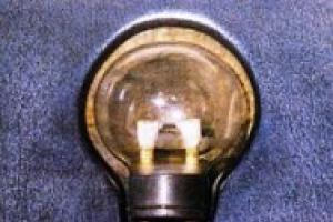
From the beginning of the last century, the twentieth century until the 60s, glow-in-the-dark radium paint (the effect of radioluminescence of the light composition, based on the reaction of 226Ra with copper and zinc) was applied to dials and hands of wall and wristwatch, alarm clocks, and were also used to coat jewelry, souvenirs, and even children’s toys and Christmas tree decorations with phosphor. Radium-226 was widely used in military equipment, in compasses and weapon sights - on airplanes, ships and submarines.
Level radioactive radiation, in the immediate vicinity of the luminous surfaces of these antique antiques, could reach large values - hundreds (for some specimens - thousands) microroentgens per hour (since, in addition to alpha particles, the 226Ra isotope also emits gamma rays with an energy of 0.2 MeV) , and approaches background values - at a distance of 1-2 meters from the source (the effect of scattering gamma rays with low energy). The usual color of luminous radium paint is yellowish or cream. The brightness of the glow, a year or two after application, noticeably decreases (zinc sulphide gradually decomposes, “burns out,” but the radiation remains, because the half-life of 226Ra is long, more than one and a half thousand years, with a bad bouquet of “daughter” isotopes) . Radium226, by chemical structure, is an analogue of calcium and when its molecules enter the human body, it can accumulate in the bones, causing internal irradiation of the body.
Until the 1930s, when in Europe they realized the dangers and consequences of exposure to strong radiation on human health, long-lived isotopes were added there to food, cosmetics and hygiene products. Due to the very high price of radium, the scale and scope of its use for civilian purposes were limited.
In modern industrial safe (if the seal of the device is not broken) permanent light compositions (SPD) with short-range sources of radioactive radiation, a mixture of radiothorium (alpha particles) and mesothorium or tritium / promethium-147 (pure beta) phosphor is used.
Radiation dose accumulates in the body in the form of irreversible changes in tissues and organs (especially intensively when high levels penetrating radiation and receiving large doses from it) and radionuclides settling in bones and tissues, causing internal irradiation (radioactive cesium-137 and strontium-90 have a half-life of about 30 years, iodine-131 - 8 days).
A level that can have a noticeable harmful effect on human health is more than 10 millisieverts per day.
Having received a radiation dose of 5 sieverts for several hours in a row, a person can die within a few weeks.
Intervention levels: to begin temporary resettlement of the population - 30 mSv per month, to end - 10 mSv per month. If the dose accumulated over one month is predicted to remain above these levels for a year, the issue of relocation to permanent residence should be considered.
With increased accuracy, you can measure radiation with a household dosimeter-radiometer by taking quite a lot of measurements at a point (at a height of 1 meter from the ground surface) and calculating the average value or with several working devices at once, followed by averaging the measurement results. Record the readings taken, the time and number of measurements, the name, model and serial number of the equipment used, as well as the location and reason for the test. If it is raining, you must indicate this, since high humidity negatively affects the operation of these devices. Visually draw a map-scheme of the gamma survey - in the form of a picture or drawing with the main elements of the situation (lines) and indicating the compass orientation at the survey site. If local foci of gamma radiation are detected with a dose rate exceeding twice the natural background for a given area, it is necessary to carefully delineate them using measurements on a ten-meter coordinate grid and contact the local SES (sanitary and epidemiological station).
Natural, terrestrial sources of increased radioactive background - due mainly to the characteristics geological structure specific area and are usually associated with nearby granite (and other intrusive rocks) massifs and flooded tectonic faults (a source of radiative emanations of radon gas from groundwater). In underground cavities, in caves and adits located there, there may be increased background radiation values, which speleologists and diggers need to take into account (you must have at least one working normal dosimeter-radiometer per group, with an audible alarm turned on).
The results of individual monitoring of personnel radiation doses must be stored for 50 years. When conducting individual monitoring, it is necessary to keep records of the annual effective and equivalent doses, the effective dose for 5 consecutive years, as well as the total accumulated dose for the entire period of professional work.
In Chernobyl, during the accident, the liquidators worked until they reached a dose of 25 rem, that is, twenty-five roentgens (this is approximately 250 millisieverts), after which they were sent from there. Health status was also monitored using regular blood tests.
There is no radiation from a cell phone, but there is electromagnetic microwave radiation (the highest power at the antenna - in talk mode and with poor quality of the received signal), which is non-ionizing, but still has a damaging effect on biological tissues, especially on the central nervous system ( on the brain) and on the state of health in general, IF you do not use a wired headset or hands free telephone headphones. Medical studies have shown that from the electromagnetic field of a telephone handset, memory deteriorates, a person’s intellectual abilities decrease, headaches and night insomnia occur. If calls on a mobile phone last more than 1 hour a day ( professional level exposure) - you must be regularly (every year) seen by a doctor (a therapist, if necessary, an oncologist). You can protect yourself if, when using headphones, you hold the mobile phone handset at a sufficient distance to reduce its radiation - no closer than half a meter from your head.
Persons exposed to a single dose of radiation exceeding 100 mSv should not be exposed to doses exceeding 20 mSv/year in further work. These people are not contagious. The danger comes from radioactive substances, for example, in the form of dust on work uniforms and the soles of shoes.
In case of emergency ( emergency), to monitor the situation - have with you an individual dosimeter (always on in accumulation mode) or a radiometer configured to sound the threshold radiation value, for example - 0.7 µSv/h (µSv/h, uSv/h - designation on English language) = 70 micro-roentgens/hour. Gas masks used in the zone of radiation contamination (especially their filters) are a source of radiation.
When coal is burned, potassium-40, uranium-238 and thorium-232 contained in it are released in microscopic quantities. For this reason, furnaces that were fired with coal, ash dumps and nearby areas over which dust and ash fell from coal smoke have some radioactivity, usually not exceeding permissible standards. Using a radiometer and a magnetometer, archaeologists find ancient sites and human dwellings located at great depths from the surface of the earth.
After the Chernobyl accident, in the “luminous” territories adjacent to the disaster site, in populated areas which were covered by a radioactive cloud - special mechanized units carried out the liquidation and burial or decontamination of buildings and property, contaminated equipment (trucks and cars, earthmoving and road construction machines). As a result of the accident, water bodies, pastures, forests and arable lands were exposed to radiation contamination, some of which are still “ringing” to this day.
From the literature, a tragic incident is known that occurred in the last century in Kramatorsk (Ukraine), when a source of Cs was lost in a crushed stone quarry. Subsequently, it was discovered in the wall of a built residential building.
Tumor (cancerous) cells can withstand irradiation up to several thousand roentgens, but healthy tissues do not survive and die at an absorbed dose of 100-400 R
Iodine-containing preparations and seafood (seaweed / Kelp) should be taken in advance, in reasonable quantities and according to the instructions - to prevent thyroid cancer from radioactive 131 I. You cannot drink a regular alcohol solution of iodine. You can only smear it externally - in the form of an iodine net (or “flowered”, under Khokhloma), draw it on the skin of the neck or other parts of the body (if there is no allergy to it).
There are several main ways to protect against penetrating radiation: limiting the exposure time, reducing the activity and energy of the radiation source, distance - the dose rate decreases with the square of the distance from the isotope (this rule only applies to small, “point sources”, relatively small linear sizes). When large areas and territories on the surface of the Earth are contaminated or when radionuclides, in the form of fine particles, enter the upper layers of the atmosphere, the stratosphere (with a sufficiently large power of nuclear warheads - from one hundred kilotons and above) - the level of radioactive radiation will be higher, the damage to the environment and danger to the population, radiation (dose) load is greater. In the event of a large-scale nuclear war, with the use of hundreds or several thousand nuclear warheads (including high and ultra-high power), in addition to radiation, there will be catastrophic consequences in the form of global (planetary scale) climate changes, abnormally cold, nuclear winter and night (lasting up to several years) - without sunlight(access solar energy will decrease hundreds of times, with a widespread decrease in air temperature by 30-40 degrees), with famine and mass extinction of the population of entire continents, the disappearance of most flora and fauna, the destruction of ecosystems, the loss of the ozone layer (which protects the Earth from destructive cosmic rays) by the atmosphere of the planet. Numerous nuclear power plants, nuclear waste storage facilities, gushing oil wells and burning gas flares, warehouses, factories and chemicals were left unattended and unmaintained after the global cataclysm. factories will add to the environmental problems of a depopulated planet. In the slang of "survivalists", such future events are called BP (from the abbreviation of the name "Big and Fluffy Northern Animal"), and before it was called the Apocalypse. Then, after the deposition of raised dust and ash on the earth and snow surfaces, when they are heated by solar radiation, a “nuclear summer” will begin, with the melting of the glaciers of the Himalayas, Greenland, Antarctica and the snow caps of the mountains, with an increase in the level of the world ocean, inland seas and reservoirs , the “global flood” will happen again. Perhaps people who took refuge in mountain caves and mines or in deep underground bunkers and shelters with a supply of food for several years, with a reserve fresh water, with air storage and regeneration systems. The opportunity to survive when the poles change will also be available to submariners of nuclear submarines who went to sea shortly before the disaster. City residents will try, for a while, to take refuge in old, unflooded bomb shelters or in city metro tunnels, while at the nearest prod. warehouses will not run out of food and drinking water. Humanity still has a chance to avoid the next and most destructive world war if they appear and optimally begin to be introduced into daily life new NBIC technologies (nano-, bio-, information and cognitive), solving civilizational problems with energy resources and food supply for the planet's population.
Oil field studies show a marked increase in radiation levels in the area of oil wells, caused by the gradual deposition of radium-226, thorium-232 and potassium-40 salts on equipment and adjacent soil. Therefore, spent oilfield drill pipes often become radioactive waste.
Non-ionizing radiation, due to its lower energy compared to ionizing radiation, is not capable of breaking the chemical bonds of molecules. But, with long-term exposure (duration) of exposure and some of its parameters (intensity, combination of frequencies, modulation of the signal and its strength, frequency of exposure) - they can adversely affect a living organism and worsen the health of people. According to the usual classification, non-ionizing radiation includes: electromagnetic radiation (in the range of industrial and radio frequencies), electrostatic field, laser radiation, constant and, especially, alternating magnetic fields (the magnitude of which is more than 0.2 µT). In modern urban conditions, human life is constantly surrounded by various non-ionizing radiation from household appliances (microwave ovens and other electrical appliances), transport, power lines, etc. They pose a danger to people with weakened immune systems, patients with diseases of the central nervous, hormonal, and cardiovascular systems. The population can be protected using various protective equipment and organizational and technical measures - limiting the time and intensity of exposure, distance (distance to the emitter) and location, using grounded protective screens (sheet metal, foil or mesh, various films and textile fabrics with a metallized coating) to weaken the fields.
Living organisms are constantly exposed to irradiation from natural sources, which include cosmic radiation, radionuclides of cosmic and terrestrial origin - 40 K, 238 U, 232 Th and their daughter nuclides, including 222 Rn (radon).
A radiologist, if he is a competent and adequate specialist, will try to minimize the total dose load for the patient so that treatment, X-ray and other examinations do not cause significant side effects for human health. But a large accumulated dose is possible if, for example, a surgeon or other doctor sends you to do x-rays many times. In order to make a correct diagnosis, this procedure can be repeated many times, and even in two or three projections.
In practice, to quickly check food products or building materials, soil and soil with a household radiometer, the filter cover is removed and the device operates ("counts") in the "indicator of excesses above the natural background" mode of gamma + hard betta radiation (if with a cover, it will measure only the gamut). To protect from water and dampness, place the device in transparent cellophane. Alpha particles cannot be detected by any household device; this requires professional equipment.
The equivalent dose rate of man-made radiation = the result of measurement by a radiometer (in microsieverts) minus the natural background radiation. In places where members of the public are located, it should not exceed 0.12 μSv/hour. For example, the background (that is, usual) value in a given area is 0.10 μSv/h, and measured there, at the outer surface of an object, is 0.15 μSv/h. Then: 0.15 - 0.10 = 0.05, which is not higher than the permissible twelve hundredths of a microsievert. This means that at this point there is no excess of 0.12 μSv/hour above the background level - technogenic radiation is “normal for the population”, in terms of radiation.
In the simplest homemade radiometer, the sensor is elongated sheets of thin newsprint or foil petals. They are attached to a metal rod placed in a glass jar. From the side, through the glass, such an indicator reacts to gamma, and if you bring an object from above, it also reacts to beta and alpha radiation (at a distance of up to 9 cm, directly, since alpha is absorbed even by a sheet of paper and a ten-centimeter layer of air). Electrify the detector static electricity it is necessary so that the time of complete discharge is at least 30 seconds, according to a stopwatch (only if the transition process is sufficiently long - the accuracy of measurements is ensured). To do this, you can use a regular plastic comb. Start and end measurements with any device, not just homemade ones, by determining the background values (if everything was done correctly, they will be approximately the same). To reduce the air humidity in the jar (so that the electroscope holds a charge) - heat it and place granules of silica gel or aluminum gel inside (pre-dry them, bake them on some fairly hot surface, in a frying pan).
// When searching for the first ones uranium deposits, for the defense purposes of our country (potential adversaries, the Americans, were already testing their nuclear weapons at that time, and their plans were to use them against the USSR), Soviet geologists also used such first sensors, in the absence of others (before measurements, the jar was dried in a hot Russian furnace) to check the level of radioactivity of the found ore samples.
An example of measurements with a homemade petal radiometer on building materials:
background value - 42 seconds (based on the results of several measurements, background = (41+43+42) / 3 = 42 s.
quartz sand - 43 pp.
red brick - 32 pp.
crushed granite - 15 s.
RESULT: the crushed stone seems to be radioactive - its radiation is almost three times (42: 15 = 2.8) higher than the background (the value is not absolute, relative, but a multiple of the background values is a fairly reliable indicator). If measurements by specialists using a professional instrument confirm the result (three times the background), the local SES (sanitary and epidemiological station) and the Ministry of Emergency Situations will take care of the problem. They will conduct a detailed radiometric survey of the contaminated area and the surrounding area and, if necessary, decontaminate the area.
Lead poisoning (Saturnism)
Heavy metals include those whose density is greater than that of iron (lead, arsenic, cadmium, mercury, cobalt, nickel). Accumulating in the human body, they cause carcinogenic effects.
Let's consider this using the example of lead (lat. Plumbum).
Lead enters the body in different ways: through the respiratory system (in the form of dust, aerosols and vapors), with food (5-10% is absorbed in the gastrointestinal tract) and through the skin. Lead compounds are soluble in gastric juice and other body fluids.
Forms of “saturnism” are weakness, anemia (pallor), intestinal colic (intestinal paralysis), nervous disorders and joint pain. One of the main signs of the disease is anemia. Brain lesions are clinically accompanied by convulsions and delirium, sometimes leading to drowsiness and coma. Of the peripheral nerves, the motor nerves are most often affected; paresis and paralysis often develop in the extensors of the hands and shoulder girdle. A gray “lead border” forms on the gums.
Lead accumulates in bones (the half-life of bone tissue is more than 20 years), nails and hair, as well as in the tissues of the liver and kidneys.
Lead encephalopathy is an acute disorder observed more often in children who have ingested lead paint. It begins with convulsions, after increased intracranial pressure and cerebral edema.
Dyes containing lead: lead white (lead carbonate, poisonous), red lead and litharge (red oxides), massicot (yellow). Enameled dishes coated on the inside with red or yellow enamel, as well as those with chips and cracks in the enamel, are harmful to health (poisoning with lead, cadmium, nickel, copper, chromium, manganese and other metals is possible).
In nature, lead ore appears as a result of the transformation of radioactive isotopes of uranium and thorium into stable (non-radioactive) isotopes of Pb with the release of alpha particles (helium nuclei).
Historical information: in 1697, the German physician Eberhard Gockel published a book entitled “A Remarkable Account of the Previously Unknown “Wine Sickness” Caused in the Years 1694, 95 and 96 by the Sweetening of Sour Wine with Lead Lithe...”, based on the results of his medical practice .
Background radiation is the level of quantum fluxes and elementary particles in the environment. This concept is important for humans when it comes to ionizing radiation. IN large quantities it poses a serious danger to living organisms. If the natural background radiation (NBR) of an area does not exceed permissible standards, then you can live on it, farm and eat the gifts of nature. When ERF is elevated, you cannot be in such places; even if you follow safety measures, you should reduce the time you spend in the contaminated area to a minimum. In some cases, radiation benefits humans. With its help, very successful treatment of cancer is carried out. The effect of isotopes on plants, insects and animals makes it possible to develop new species that differ in a set of positive properties.
Types of radiation
The natural radiation background is influenced by the number of elementary particles that previously hit the area or object and continue to come from various sources.
Modern science distinguishes between types of radiation that directly affect the natural radiation background:
- Gamma radiation. It is a flow of microparticles with a neutral charge. Has high penetrating ability. This type of radiation is the most destructive to all living things. Materials with heavy nuclei provide protection against X-rays. They trap gamma particles, becoming a source of radiation.
- Beta radiation. Its carrier is larger particles with average penetrating ability. Potentially dangerous to humans, beta rays are trapped in a thin layer of metal, wood and stone.
- Alpha radiation. It is a stream of heavy positively charged particles. They carry a powerful ionic charge that has a destructive effect on living tissue cells. In humans, alpha particles only affect the outer layer of the skin. Even clothing is a barrier for them.
On earth, sources of radiation that create natural and artificial background radiation are the sun, stars, rocks and industrial facilities built by man. The level of contamination is created by isotopes of chemical elements such as iodine, uranium, radium, strontium, cobalt, cesium and plutonium. Knowing what radiation is, you can successfully protect yourself from such a phenomenon dangerous to life and health.
Sources of natural radiation
Until the Earth acquired an iron core and received an impulse to rotate, it was open to all types of radioactive radiation. After a powerful magnetic field formed around our planet, it gained protection from penetrating radiation. The solar wind, destructive for all living things, bends around the Earth along the magnetic field lines. A small portion of heavy alpha particles hits the planet's surface. They pose a danger only when exposed to the sun for a long time without protection. This causes skin burns.
The volumetric energy emissions produced by pulsars pose a certain danger. These space objects in one second they produce as much energy as the Sun produces in a thousand years. From such a ray earth's atmosphere doesn't save.
The terrain and soil composition have a certain influence on the formation of background radiation. The most ancient rock, formed billions of years ago, is granite. Where this mineral comes to the surface or is under a thin layer of soil, there is an increased level of radiation.
Radiation levels are also affected by altitude. With every kilometer of rise above the ground, the thickness of the protective layer of the atmosphere decreases. Already at an altitude of 10,000 meters there is such a radiation background, the norm of which is close to the maximum permissible.
Depending on the geographical location The radiation level changes. At the poles it is much stronger than at the equator. This phenomenon is caused by the shape of the Earth's magnetic field, which converges at the poles.
Soil characteristics. The highest levels of radiation are observed in places where uranium ore occurs. Even if the deposit of this chemical element is located several kilometers underground, the level of its radiation can exceed the maximum permissible by several times. Iron ore and bauxite can create a small background. These elements tend to accumulate radiation.
Artificial radiation on earth
This phenomenon is an excess of the natural background due to human activity. The history of the development of the atom goes back several decades. Since this area of industry has not yet been fully developed, the risk of emergency situations is quite high.
Background radiation standards may be exceeded for the following reasons:
- Conducting nuclear weapons tests. The area where atomic bomb tests were carried out is saturated with radioactive isotopes. It will be uninhabitable for many centuries to come.
- Use of the atom for peaceful purposes. Nuclear charges were used to change the course of rivers, create artificial reservoirs, and to extinguish fires in gas fields.
- Accidents at nuclear power facilities. During such incidents, isotopes are released into the atmosphere. Depending on the scale of the accident, the surrounding area becomes uninhabitable for a period of 30 to 10,000 years.
- Accidents during transportation and disposal of nuclear fuel and waste. As a result, material contaminated with isotopes is spread over a wide area.
Depending on the degree of radioactive contamination of the area, stay on it may be limited in time or prohibited completely.
Consequences of radioactive contamination
The level of radiation is measured in the number of isotopes received per unit of time. The radiation power is determined in roentgens per hour, the dose received is calculated by summing up all indicators for the year. This component is measured in grays (Gy).
Depending on the volume of isotopes absorbed by the body, a person can get radiation sickness:
- I degree. The disease does not pose a danger to humans provided they are evacuated from the contaminated area. It manifests itself in the form of weakness, headache, sleep and appetite disturbances. When receiving a dose of up to 2 Gy, recovery can occur within one and a half to two months.
- II degree. If a dose of up to 4 Gy is received, moderate damage occurs. The patient experiences acute pain, the activity of internal organs and the central nervous system is disrupted. Externally, the disease manifests itself as hair loss, teeth loss and the formation of ulcers. Even qualified treatment does not provide complete recovery.
- III degree. A dose of 4-6 Gy causes irreversible processes in the human body. Severe disease leads to failure of internal organs and necrosis of soft tissues. As a rule, with concomitant loss of immunity, the disease is fatal.
- IV degree. A severe form develops when the patient receives more than 6 Gy. It is not possible to describe the symptoms experienced by the patients, since their death occurred within a matter of hours after exposure. The death was preceded by complete destruction of the soft tissue structure, cardiac arrest and cessation of breathing.
Radiation injury is considered to be a person receiving a dose of less than 1 Gy.
Current background radiation standards
Radiation standards are averaged, obtained from the results of clinical studies of patients who received radiation doses of various levels. People can receive the resulting total doses over different periods of time. The greater the radiation intensity, the more dangerous the consequences and the more difficult the treatment. Therefore, the definition of what normal background radiation is is established at the legislative level and is a value for regulating living or working conditions at an enterprise.
Radiation safety rules apply to the following categories of citizens:
- military personnel serving on nuclear submarines and surface ships;
- NPP personnel;
- people living in areas with high background radiation;
- professional rescuers and emergency crew workers working at nuclear energy facilities;
- medical workers who deal with devices containing radioactive elements;
- scientists working with radioactive material.
According to studies, radiation with a power of 20 microroentgens per hour is considered absolutely safe for the health of an adult.
The radiation limit is considered to be 50 microroentgens per hour. However, if over the course of a year, receiving small doses of radiation at regular intervals, a person receives a total of 1 roentgen, then it will be practically safe for him. Radiation is gradually eliminated from the body. The radioactive safety standards in force today determine the maximum dose of radiation received over a lifetime within 60-70 roentgens.
If we take the level of exposure to background radiation and gamma radiation in microsieverts per hour, then the acceptable safety limit is considered to be:
- watching TV 3 hours a day for a year (0.005 mSv);
- long flight by plane (0.01 mSv);
- being in an open area in sunny weather (1 mSv);
- work at nuclear power plants (0.05 mSv).
A dose of 11 μSv per hour is considered dangerous. It increases the risk of cancer.
Radiation in a general sense refers to the spread of energy in the form of elementary particles and quantum fluxes. There are light (visible to the naked eye), infrared, ultraviolet and ionizing radiation.
For the safety of human life, the greatest interest is ionizing radiation, which promotes the formation of free radicals in the cells of a living organism, which triggers the process of protein destruction, cell death or degeneration.
These processes can cause the death of a living organism. That is why the term “radiation” most often means ionizing radiation.
Are all types of radiation dangerous?
Radiation exposure is not always fatal and destructive, as is commonly believed. In some cases, the instability of isotopes of various elements is used for good, in particular, in plant and animal breeding, medicine, energy and the national economy.
Are radiation and radioactivity the same thing?
Radiation and radioactivity are similar concepts, but not at all identical. Radiation is the name given to free flows of energy that exist in space until they are absorbed by some object. Radioactivity is the ability of an object or substance to absorb radiation, becoming a source of radiation.
Types of radiation and penetrating power
There are several types of radiation, among the most significant are the following:
- Alpha radiation is a stream of positive particles with a relatively large mass; they have powerful ionization and pose a serious danger when entering the body through the gastrointestinal tract, but are retained even by small barriers and do not penetrate the skin.
- Beta radiation is tiny particles with slightly greater penetrating power. A thin layer of aluminum or a few centimeters of wood can protect against such radiation.
- Gamma radiation and similar X-rays - a stream of neutrally charged particles with high penetrating ability - pose the greatest danger to humans. Materials with heavy nuclei can protect against radiation, and this will require a layer of several meters.
Natural and artificial radiation
Radiation can be either natural or due to human activity. In nature, powerful sources of radiation are the Sun and the process of decay of certain elements in the composition earth's crust. Even in the human body, there are normally substances that create a personal radiation background.
Artificial radiation is a consequence of the activities of nuclear power plants, the development and use of any technology that uses nuclear reactors, as well as the use of radioactive isotopes in medicine, mining of elements with unstable atomic nuclei, testing, hazardous waste disposal and nuclear fuel leaks.
External and internal exposure
Natural background radiation is determined by the presence of external and internal sources of radiation. The main ways radiation enters the human body are:
- through the digestive tract, which is determined by living conditions and the nature of human activity;
- through mucous membranes and skin, which is also determined by location and may be related to the characteristics of the area of residence (affected by the proximity of artificial sources of radiation, geographic latitude and altitude above sea level) and building materials, containing radioactive substances from which housing and infrastructure facilities are built.
Permissible and lethal doses of radiation
The natural level of radiation depends on the area and human living conditions. The value is measured in doses received by the body over a certain period of time (usually one hour or a year):
- Exposure, reflecting the degree of ionization with gamma or x-ray radiation, the basic unit of measurement is the x-ray.
- The dose absorbed by a substance, object or organism is measured in “grays”.
- The effective (permissible) dose is determined individually for each organ.
- The equivalent dose of radiation exposure is calculated according to coefficients and depends on the type of radiation.
Radiation standards
On average, the normal radiation value and does not pose a danger to the population is about twenty microroentgens per hour, but the figure can vary significantly depending on the characteristics of the territory under study.
The maximum radiation limit (MPC - maximum permissible concentration) is an indicator of approximately 0.5 μSv/hour (or 50 μR/hour). However, when the duration of exposure to radioactive radiation is reduced to several hours, a person can endure radiation doses as high as 10 μSv/h (or 1 μR per hour).
Being in an area of radiation contamination or exposure to radiation, for example, when medical research, a few minutes the maximum permissible level of exposure is up to several millisieverts per hour.
Penetrating radiation accumulates in the body. The standards determine that for the full functioning of the body and maintaining health at the proper level, the accumulated amount of radiation over a lifetime should not exceed a limit of 100 to 700 mSv.
At the same time, in the field of upper values, permissible doses will be for residents of high mountain areas and territories with increased radioactivity.
A table of approximate radiation doses at various types activities. For example, with fluorography the dose received is 0.06 mSv, and x-ray gives 30% and 3% of radiation exposure from the annual dose for X-rays (film and digital, respectively) of the chest organs.

Radiation contamination
Radiation (radioactive) contamination is a situation that poses a danger to the health and even the lives of people living in areas where radioactive substances fall out, as well as in areas close to the epicenter of man-made accidents. Normal background radiation is disrupted by leaks during transportation and storage of radioactive waste, accidents at nuclear power plants, or as a result of accidental or intentional loss of radio sources.
The main toxic substances are iodine-131, strontium, cesium, cobalt and americium. The minimum half-life of radioactive substances is about eight days, the maximum is more than four hundred years. In case of man-made accidents, radiation doses are reduced to an acceptable level on average within 30-50 years, although everything depends on the nature of the release.
For example, being in the exclusion zone around the Chernobyl nuclear power plant for 10 hours today is equivalent to flying, and in Hiroshima and Nagasaki, which experienced the impact nuclear bomb, people can live at the moment.
Dangerous doses of radiation
- A 50% probability of death occurs with 3-4 Gy of penetrating radiation, and with 7 Gy or more, death occurs in 99% of cases;
- Irradiation above 10 Gy can already be considered fatal for a person; radiation sickness in this case kills in 2-3 weeks.
- The lethal dose of radiation for humans is 15 Gy (death occurs within 1-5 days);
Symptoms and severity of infection
The clinical picture of radiation sickness is divided into four degrees of severity:
- first degree damage occurs with irradiation within 2 Gy;
- moderate severity is typical for doses up to 4 Gy;
- in severe (third) degree, radiation ranges from 4-6 Gy;
- The radiation dose for extreme radiation sickness is more than 6 Gy.
In addition, doctors talk about radiation injury occurring without any characteristic symptoms if the victim received radiation less than 1 Gy.
- Symptoms of the first degree of radiation sickness include headaches, changes in appetite, irritability and sleep disturbances. Victims usually experience irritation of the mucous membranes, gastrointestinal disorders and increased sweating. Recovery occurs within one to two months if exposure to radiation has stopped.
- Defeat medium degree severity is characterized by aggravation of existing symptoms, pathological changes in internal organs and the central nervous system, the occurrence of trophic ulcers, as well as numerous complications that are associated with weakened immunity. Patients often never fully recover, and doctors only manage to achieve remission with periodic exacerbations.
- Radiation sickness of the third degree is characterized by irreversible changes in the functioning of most organs and systems, tissue degradation and frequent bleeding. The condition poses a significant threat to the patient's life, progresses rapidly and in most cases ends in death.
- Signs of radiation damage of extreme severity have been little studied in medical practice, because Such a serious form of radiation sickness is very rare. Modern methods diagnostics and treatment make it possible to identify and stop the disease at those stages when it is still advisable to provide assistance to the victim. In this case, a persistent improvement in the patient’s condition occurs, as a rule, two to three years after the cessation of exposure to radiation on the body.

